
Recently, Professor Yin Chenghong's team from Beijing Obstetrics AND Gynecology Hospital Affiliated to Capital Medical University published a paper entitled "SESN2 suppresses ferroptosis in polycystic ovary syndrome by maintaining PRDX6 K209 lactylation" in the journal "FREE RADICAL BIOLOGY AND MEDICINE", a journal in the Q1 area of biology. This study proposed for the first time a new regulatory axis of "SESN2-PRDX6 lactation modification" and elucidated the molecular mechanism by which PRDX6 expression regulates ovarian function.
Polycystic ovary syndrome (PCOS) affects 8-10% of women of childbearing age worldwide and is the leading cause of ovulation-dysfunctional infertility. Recently, a study published in Free Radical Biology and Medicine revealed a new "SESN2-PRDX6 lactylate" signaling axis, which acts like an "invisible guardian" to inhibit ferroptosis in ovarian granulosa cells through a protein modification called "lactoylation", thereby protecting ovarian function. This discovery opens up a promising new direction for the treatment of PCOS.
New "lethal" switch in PCOS: ferroptosis and protective effects of SESN2
Ferroptosis is an iron-dependent, programmed cell death mode characterized by lipid peroxidation, which has been proven to be a key driver of follicular dysplasia and ovulation abnormalities in PCOS patients in recent years. The study found that ferroptosis was significantly activated in PCOS model mice and cells. A stress-induced metabolic regulator called Sestrin 2 (SESN2) plays a key role in this.
The research team systematically validated this conclusion by constructing two independent PCOS mouse models (dehydroepiandrosterone DHEA-induced model and letrozole-induced insulin resistance PCOS model) and a human granulosa cell line (KGN) model. In DHEA-treated human KGN granulosa cells, cell viability decreased, while SESN2 expression levels increased compensatorically (Figure 1B-D). However, when SESN2 expression was knocked down using small interfering RNA, the situation worsened dramatically: cell viability was further reduced compared to the DHEA-treated group alone (Figure 1G), indicating that endogenous SESN2 is essential for maintaining the survival of granulosa cells under stress.
Further mechanistic studies showed that the deletion of SESN2 exacerbates oxidative stress and ferroptosis. Specifically, DHEA treatment could cause an increase in mitochondrial reactive oxygen species levels, while the level of mitochondrial superoxides further increased after SESN2 was knocked down. At the same time, the antioxidant "gold standard" in cells, glutathione content and superoxide dismutase activity, are also disturbed. Notably, although DHEA treatment increased glutathione levels (possibly a compensatory response), this effect was eliminated after SESN2 knockdown and superoxide dismutase activity was further reduced (Figure 1H-I). Lipid peroxide probe C11-BODIPY staining directly showed a significant increase in green fluorescence intensity, which represents lipid oxidation, after SESN2 deletion (Figure 1L). These changes can be partially reversed by ferroptosis-specific inhibitors Ferrostatin-1 or iron chelator deirarox.
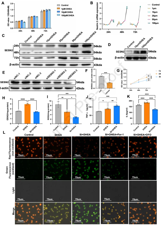
Figure 1
Key mechanism: How SESN2 maintains K209 lactylation of PRDX6
So, what "molecular switch" does SESN2 play a protective role? The answer lies in a subtle protein post-translational modification – lysine lactacylation.
The researchers performed lactylate modification omics analysis on normal and SESN2-knockdown KGN cells. The results were striking: SESN2 deletion led to a significant decrease in cell-wide lactylate levels, with 2,108 lactacylation sites significantly downregulated on 988 proteins and 145 loci upregulated on only 106 proteins (Figure 3B). Among the many differentially modified proteins, an antioxidant enzyme called Peroxiredoxin 6 (PRDX6) caught the team's attention. PRDX6, a key intracellular phospholipid hydroperoxide reductase, has lysine lactylation at position 209 significantly reduced after SESN2 deletion (Figure 3D).
Further immunoprecipitation assays confirmed that PRDX6 has a direct physical interaction with glutathione peroxidase 4, a core inhibitory protein of ferroptosis (Figure 3H). More importantly, the deletion of SESN2 significantly reduced the overall lactylation level of PRDX6 (Figure 3I-J), which provided a molecular basis for subsequent loss of function.
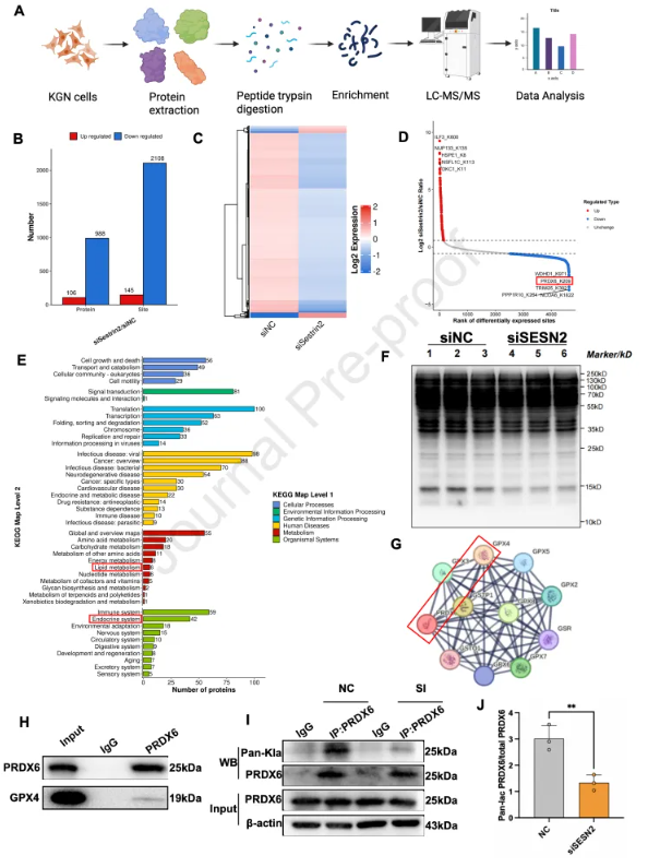
Figure 3
Preclinical validation: SESN2 deletion exacerbates ovarian dysfunction in PCOS mice
To verify the authenticity of this mechanism in vivo, the research team constructed SESN2 knockout mice and validated them in DHEA and letrozole-induced PCOS models, respectively.
In the DHEA-induced PCOS model, PCOS mice with SESN2 gene knockout were significantly worsened compared to normal PCOS mice. The estrous cycle is completely disturbed, and the number of cystic follicles in the ovarian tissue increases significantly, while the number of corpus luteum, which represents ovulation, is further reduced (Figure 5C-G). Serum hormone testing also confirmed this phenotype: testosterone levels in SESN2-knockout PCOS mice spiked further to the highest value of all groups (Figure 5H), while follicle-stimulating hormone levels decreased even lower (Figure 5J).
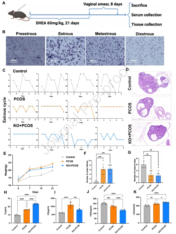
Figure 5
At the tissue level, marker 4-hydroxynonenal staining representing lipid peroxidation and Prussian blue staining representing iron deposition were significantly enhanced in PCOS ovarian tissue with SESN2 deletion (Fig. 6A-B). At the same time, the expression of key proteins in ferroptosis was also altered: the inhibitors GPX4 and FTH1 were further decreased, while the promoters ACSL4 and COX2 were further increased (Figure 6C). In the letrozole-induced PCOS model, the team observed highly consistent results that SESN2 deletion also exacerbated abnormal glucose tolerance, hormonal disturbances, and ferroptosis damage to the ovaries (Figures 7-8). Notably, SESN2 knockout was accompanied by a significant decrease in PRDX6 protein expression in both in vivo models (Figure 6E, Figure 8E).
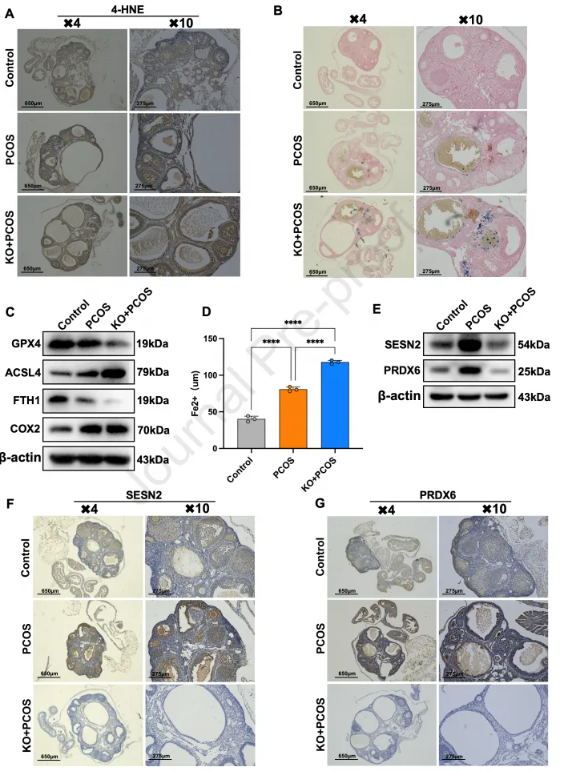
Figure 6
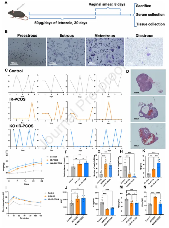
Figure 7
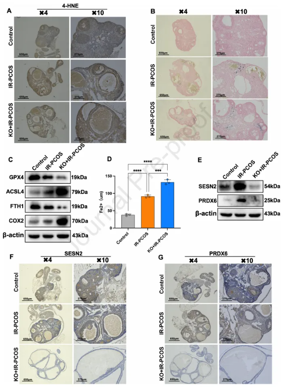
Figure 8
PRDX6 K209 lactyl is the key to resisting ferroptosis
In order to finally lock in the function of PRDX6 K209 lactylate, the research team conducted a subtle "replenishment" experiment. They were transferred to normal PRDX6 (wild-type) and K209R mutants that mimic non-lactoylation (mutating lysine to arginine and not being lactacylated) in SESN2-knocked down and DHEA-treated cells, respectively.
The results clearly showed that the K209 site of PRDX6 was the "lifeline" for its protective role. Supplementation of wild-type PRDX6 significantly restores the interaction between PRDX6 and GPX4 (Figure 4B) and effectively reverses the decrease in glutathione and superoxide dismutase levels caused by SESN2 deletion (Figure 4E-F). At the same time, lipid peroxidation (Figure 4G-H) and cell death (Figure 4I-J) were also significantly inhibited.
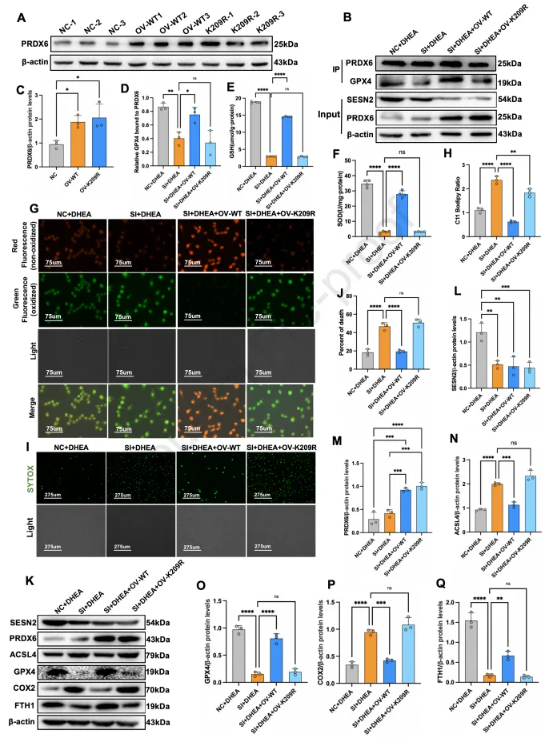
Figure 4
However, the K209R mutant was almost completely lost in this protection. It cannot effectively restore the binding of PRDX6 to GPX4, nor can it effectively inhibit lipid peroxidation and cellular ferroptosis. This result irrefutably proves that the lactylate modification of lysine at position 209 of PRDX6 is the key to its antioxidant and anti-ferroptosis functions.
This study reveals for the first time a new protective signaling pathway in PCOS: SESN2, as a stress-inducing factor, stabilizes the interaction between PRDX6 and GPX4 by maintaining the lactylate modification of the K209 position of the PRDX6 protein under stress conditions such as elevated androgens, thus forming a strong antioxidant defense network, inhibiting ferroptosis in granulosa cells and protecting ovarian function. This line of defense collapses when SESN2 function is lost or K209 lactacylation of PRDX6 is blocked, leading to oxidative damage and increased ferroptosis, ultimately contributing to follicular dysplasia in PCOS.
The graphical summary of the study clearly summarizes this mechanism: androgen stimulation such as DHEA induces oxidative and ferroptosis stress while triggering compensatory elevation of SESN2. SESN2 acts as an adaptogenic protective factor to help maintain the lactylate of PRDX6 and the antioxidant axis of PRDX6-GPX4. However, this endogenous response is not sufficient to completely counteract the damage, and the deletion of SESN2 will further impair this protective pathway, thereby exacerbating lipid peroxidation and ferroptosis.
This study elucidates the important role of protein lactation modification in reproductive endocrine diseases, and points out that the K209 lactylate site of SESN2 and PRDX6 may be a potential new target for PCOS treatment. In the future, up-regulation of SESN2 expression by drug or gene means, or directly mimicking the K209 lactation of PRDX6, may be an effective strategy to combat ovarian function decline in PCOS patients. Of course, the researchers also pointed out that there is still a lack of direct verification of PRDX6 K209 lactyylation in vivo, and its precise biochemical consequences still need to be explored in depth, but this discovery undoubtedly lights up a new light for the treatment of PCOS.
source:Ying-ying Li, Ning Xu, Xiao-yi Gu, Bo Yu, Ying-yi Luan, Cheng-hong Yin, SESN2 suppresses ferroptosis in polycystic ovary syndrome by maintaining PRDX6 K209 lactylation, Free Radical Biology and Medicine, Volume 251,2026, Pages 75-90, ISSN 0891-5849.
Editor-in-charge:lucy