





















Key Recommendations
- It is recommended that pregnant women attend obstetric care immediately once they perceive reduced fetal movements (RFM) or absent fetal movements. [Grade B Recommendation]
- There is insufficient evidence to recommend standardized fetal movement counting based on fixed numerical cut-off values. [Grade A Recommendation]
- All clinicians should recognize that RFM is correlated with fetal growth restriction (FGR), small for gestational age (SGA) infants, placental insufficiency and congenital fetal malformations. [Grade C Recommendation]
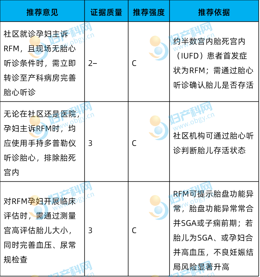
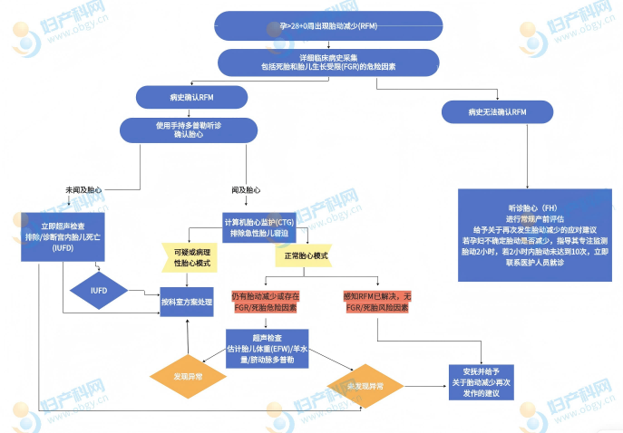
- Handheld Doppler fetal heart auscultation shall be conducted for all pregnant women with RFM presenting at community or hospital settings to prioritize exclusion of intrauterine fetal death. [Grade C Recommendation]
- Clinical assessment of women reporting RFM shall include measurement of symphysis-fundal height (if serial growth ultrasounds have not been performed regularly), blood pressure monitoring and urinalysis. [Grade C Recommendation]
- After confirming fetal viability, computerized cardiotocography (CTG) shall be performed for women with RFM at ≥26⁺⁰ weeks’ gestation to rule out acute fetal distress; diagnostic protocols for gestations below this threshold are detailed in Chapter 14. [Grade B Recommendation]
- Ultrasound examination shall be arranged for women with RFM at ≥28⁺⁰ weeks’ gestation if any of the following criteria are met: persistent RFM despite normal cardiotocography; presence of high-risk factors for FGR or stillbirth; no ultrasound screening conducted within the past 2 weeks. [Grade B Recommendation]
- Abnormal cardiotocography results shall be discussed with a senior obstetrician to formulate delivery decisions based on gestational age and the severity of tracing abnormalities. [Good Practice Point (GPP)]
- If ultrasound identifies SGA fetus, abnormal umbilical artery Doppler flow or oligohydramnios, management shall follow RCOG Green-top Guideline No.31 Screening and Management of Small for Gestational Age Infants and Fetal Growth Restriction. [Good Practice Point (GPP)]
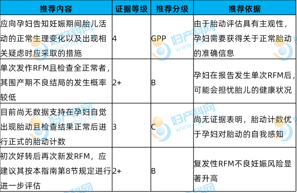
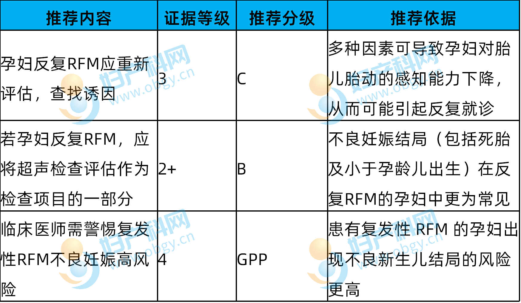
- Women with recurrent RFM after a single unremarkable workup shall be advised to return for obstetric review, with management protocols specified in Chapter 8 of this guideline. [Grade B Recommendation]
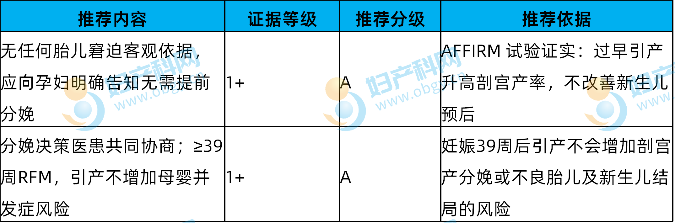
- Induction of labor is not indicated if there is no objective evidence of fetal distress (normal CTG, absence of FGR, oligohydramnios or abnormal umbilical artery Doppler). [Grade A Recommendation]
- Decisions for early delivery shall be individualized and made through shared decision-making with the pregnant woman. Elective early delivery for RFM at >39⁺⁰ weeks’ gestation is not associated with increased maternal or neonatal adverse outcomes. [Grade A Recommendation]
- Full review of clinical history shall be undertaken for women with recurrent subjective RFM to identify underlying contributory factors. [Grade C Recommendation]
- For multiple gestation pregnancies presenting with RFM, cardiotocography, fetal growth assessment, amniotic fluid volume measurement and umbilical artery Doppler ultrasound shall be completed to exclude fetal distress. [Grade C Recommendation]
Supplementary Gestation-Specific Recommendations
- RFM at 24⁺⁰–28⁺⁰ weeks’ gestation: Handheld Doppler confirmation of fetal heart sounds, clinical history taking to screen for intrauterine fetal death and high-risk factors for early-onset FGR. [Good Practice Point (GPP)]
- RFM at <24⁺⁰ weeks’ gestation: Handheld Doppler auscultation to confirm fetal cardiac activity. [Good Practice Point (GPP)]
1. Objectives and Scope of Application
1.1 Objectives
This guideline provides standardized clinical diagnostic and management pathways for community and hospital settings based on optimal available clinical evidence, to standardize care for pregnant women presenting with subjective RFM during gestation, and to summarize high-risk factors linked to RFM as well as variables affecting maternal perception of fetal movements. Due to limitations in existing research evidence, most recommendations carry low evidence grades. This guideline serves as a general practical reference for midwives and obstetricians, with individualized clinical management permissible based on each patient’s clinical condition. The term “pregnant woman” is used uniformly throughout the text.
1.2 Target Population and Clinical Settings
Pregnant women presenting with subjective RFM at community or hospital healthcare facilities.
2. Research Background
Perceived fetal movements serve as a critical marker of normal intrauterine fetal wellbeing. Most women first perceive fetal movements between 16 and 24 weeks’ gestation, with regular fetal movement patterns established from 28 to 32 weeks’ gestation. A sudden reduction or complete loss of fetal movements signals abnormal placental function and carries an increased risk of intrauterine fetal death. Multiple placental pathological studies confirm an association between RFM and organic placental lesions. Large observational clinical studies validate that abrupt, marked reduction in fetal movements constitutes a vital clinical warning sign, as corroborated by UK confidential enquiries into non-malformed term stillbirths, intrapartum perinatal deaths and multiple gestation perinatal mortality conducted in 2015 and 2017. Several studies report that a proportion of intrauterine fetal deaths are preceded by an episode of excessive, violent fetal movements. Per the Codac classification system, the UK stillbirth rate in 2022 stood at 3.35 per 1000 births; 33.9% of these stillbirths had an unknown cause, while 36.3% were attributable to placental pathology. Epidemiological data from regions implementing standardized RFM care pathways demonstrate that standardized RFM management does not reduce the overall stillbirth rate, yet women with RFM demonstrate significantly higher rates of SGA infants and adverse pregnancy outcomes. A meta-analysis incorporating 39 studies found that women with RFM had an odds ratio of 3.44 (95% CI 2.02–5.88) for stillbirth and 1.37 (95% CI 1.16–1.61) for delivery of an SGA infant.
3. Limitations of Available Data
(1) No unified global diagnostic thresholds to distinguish normal versus abnormal fetal movements exist, with heterogeneous definitions adopted across all published studies.
(2) Large-cohort (>1000 participants) descriptive studies are scarce, and randomized controlled trials investigating RFM management are limited with highly heterogeneous trial protocols, precluding pooled meta-analytic synthesis.
(3) Stillbirth represents a low-probability adverse outcome. To validate an intervention reducing stillbirth rates from 4.0‰ to 3.6‰ (10% relative reduction), a single trial arm would require enrolment of over 370,000 participants. Most existing studies suffer from insufficient sample sizes, selection bias and outcome ascertainment bias, limiting the validity of their conclusions.
4. Physiological Normal Fetal Movement Standards
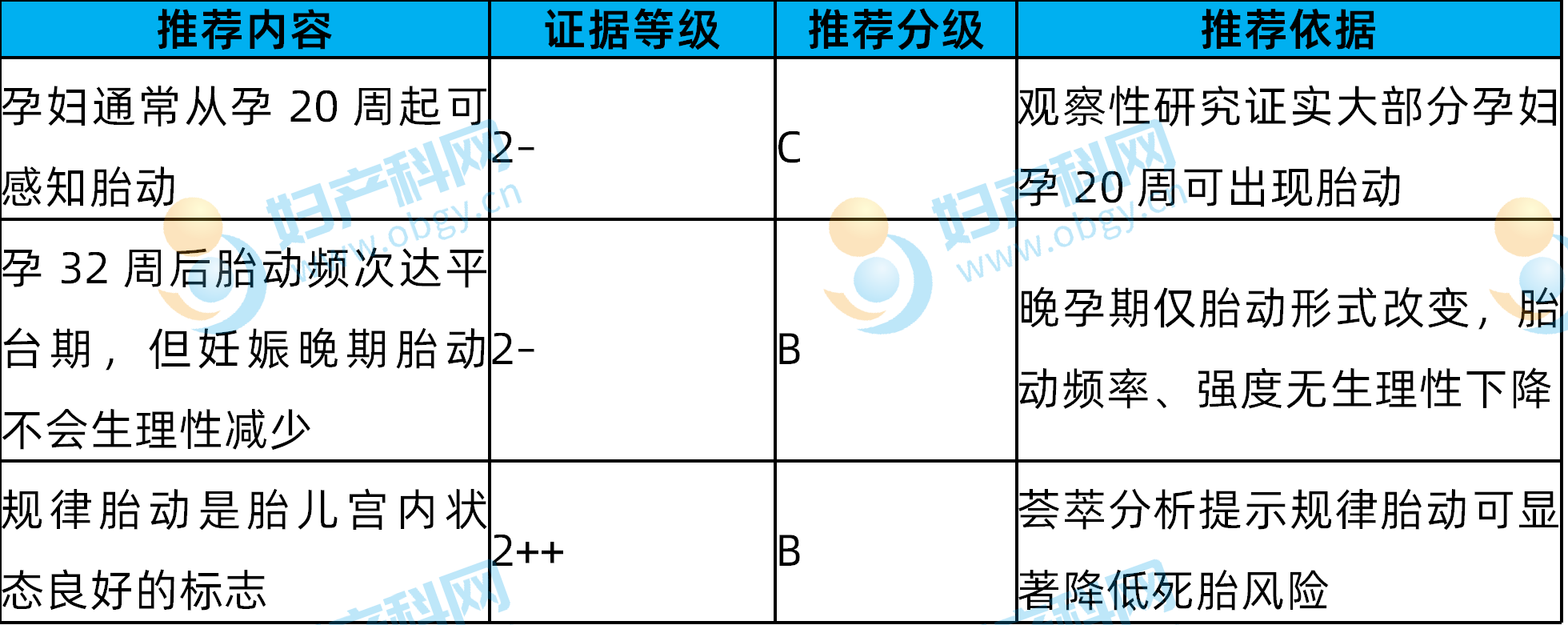
Perceived fetal movements encompass any fetal kick, flutter, sliding motion or rolling sensation felt by the mother. Fetal movements reflect intact development of the fetal central nervous, skeletal and muscular systems. Healthy fetuses exhibit spontaneous physical activity and alternate between active and sleep-wake cycles.Multiparous women may first detect fetal movements as early as 16 weeks’ gestation, while nulliparous women typically experience first perception between 18 and 20 weeks, with a subset reporting first movements after 20 weeks. Fetal movement frequency increases progressively until 32 weeks’ gestation.
- After 32 weeks’ gestation, 90% of women report unchanged or increased frequency and intensity of fetal movements until labour onset, with only a shift in movement morphology: large kicking motions gradually transition to wriggling, stretching and gentle pressure sensations.
- Term fetuses produce an average of 31 whole-body movements per hour (range 16–45), with the maximum interval between consecutive fetal movements ranging from 50 to 75 minutes.
- Fetal sleep cycles last 20–40 minutes; healthy fetuses rarely sustain sleep periods exceeding 90 minutes. Fetal movements follow a distinct circadian rhythm, with peak activity in the afternoon and evening hours.
- Increased fetal movement intensity/frequency and fetal hiccups are associated with reduced stillbirth risk (adjusted OR 0.18 and 0.42 respectively).
5. Clinical Advice on Maternal Perception of Fetal Activity
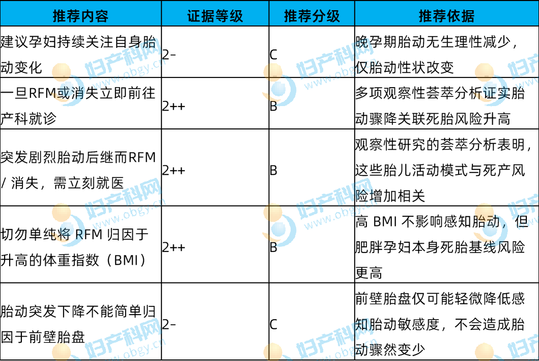
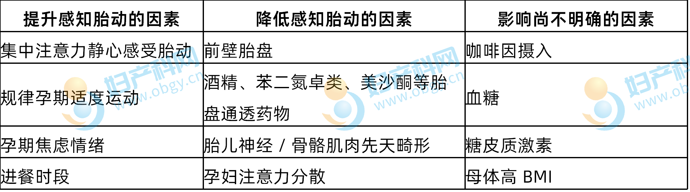
Factors Influencing Maternal Perception of Fetal Movements
(1) Perception accuracy: Concordance between ultrasound-detected fetal movements and maternal subjective perception ranges from 37% to 88%; large, sustained movements exceeding 7 seconds are most readily detected by the mother.
(2) Postural effects: Lateral positioning optimizes fetal movement perception, followed by seated posture, with standing yielding the poorest detection. Fetal activity is generally higher overnight. Women in quiet seated focus require an average of 10 minutes to perceive 10 discrete fetal movements, compared with 162 minutes during routine daily activity.
(3) Placental location: Anterior placentation may reduce movement detection before 28 weeks’ gestation, with conflicting study results after 28 weeks. Fetal anterior spinal presentation markedly diminishes maternal perception of movements.
(4) Pharmacological and environmental factors: Alcohol, benzodiazepines, opioids, glucocorticoids and carbon monoxide from tobacco smoke transiently suppress fetal activity. Caffeine alters fetal motility, though study conclusions remain inconsistent.
(5) Maternal physiological status: Both fasting and postprandial states modify fetal activity; 73.6% of women report increased movements post-meal, while 36.8% experience heightened fetal activity during fasting.
(6) Other confounding variables: Some women between 28 and 36 weeks’ gestation misinterpret uterine contractions as fetal movements. Women with a prior history of stillbirth demonstrate significantly heightened sensitivity to changes in fetal activity.Clinical Reminder: A subset of women report a pre-terminal fetal warning pattern: brief episodes of intense, agitated fetal movements followed by complete cessation of activity. This characteristic pattern is documented in 10% of intrauterine fetal death cases. Many women delay clinical presentation due to misinformation, including the misconception that fetal movements naturally decline in late pregnancy, or misguidance from lay contacts.
6. Modalities for Assessment of Fetal Movements

(1) All research protocols for objective quantification of fetal activity utilize ultrasound scanning, with single monitoring sessions limited to 20–30 minutes in semi-recumbent positioning. Ultrasound detects only 31.4%–57.2% of total fetal movements, while maternal subjective perception captures a mere 30.8% of all fetal activity.
(2) Novel monitoring devices: Abdominal accelerometers, fetal electrocardiography and fetal movement recorders remain in early-phase clinical trials. Some devices capture 82% of maternally perceived movements with an overall accuracy of 90%, yet readings are susceptible to interference from maternal coughing and abdominal wall muscular activity. Such devices are not validated for universal objective fetal movement quantification in all pregnancies.
7. Routine Standardized Fetal Movement Counting: Is It Indicated?
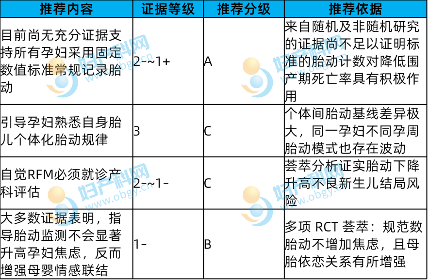
Summary of Seven Quantitative Studies Evaluating Maternal Fetal Movement Perception Interventions
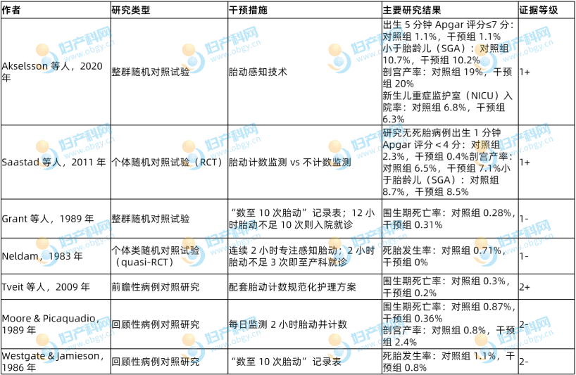
(1) Grant’s large-cohort study (68,654 participants): Participants were allocated to a standardized 10-movement counting group or routine antenatal education. Standardized fetal movement counting did not reduce perinatal mortality; overall population perinatal mortality declined from 4.0‰ pre-study to 2.9‰, attributed to general public antenatal fetal movement education.
(2) Neldam’s study (2,250 high-risk pregnancies): Weekly 3 sessions of 2-hour focused fetal movement counting versus no fetal movement counselling. Eight intrauterine fetal deaths occurred in the control arm, with zero stillbirths in the counting intervention group, demonstrating clear benefit for high-risk cohorts.
(3) Saastad (1,076 participants, “count to 10” protocol initiated after 28 weeks): The intervention group exhibited reduced rates of 1-minute Apgar <4 and improved prenatal detection of fetal growth restriction, without increased maternal anxiety.
(4) Swedish Mindfetalness multicentre trial (39,865 participants): Interventions focused on quality rather than quantity of fetal movements. No overall population reduction in stillbirth was observed, yet rates of SGA infants and caesarean delivery declined.
(5) Selected retrospective studies indicate that hospital admission upon failure to detect 10 movements within 2 hours reduces perinatal mortality, accompanied by elevated rates of hospital presentation, labour induction and emergency caesarean delivery.
8. Standardized Clinical Management of RFM
Primary clinical objectives for initial presentation: First, rule out intrauterine fetal death (incidence of IUFD among women presenting with RFM <1%); second, identify fetal distress and stratify patients at high risk of adverse pregnancy outcomes to avoid excessive medical intervention. Clinical surveys demonstrate heterogeneous awareness and inconsistent adherence to standardized RFM care pathways among global obstetricians and midwives; while clinicians recognize the association between RFM and FGR, compliance with complete diagnostic workup remains suboptimal.
8.1 Key Components of Clinical History Taking
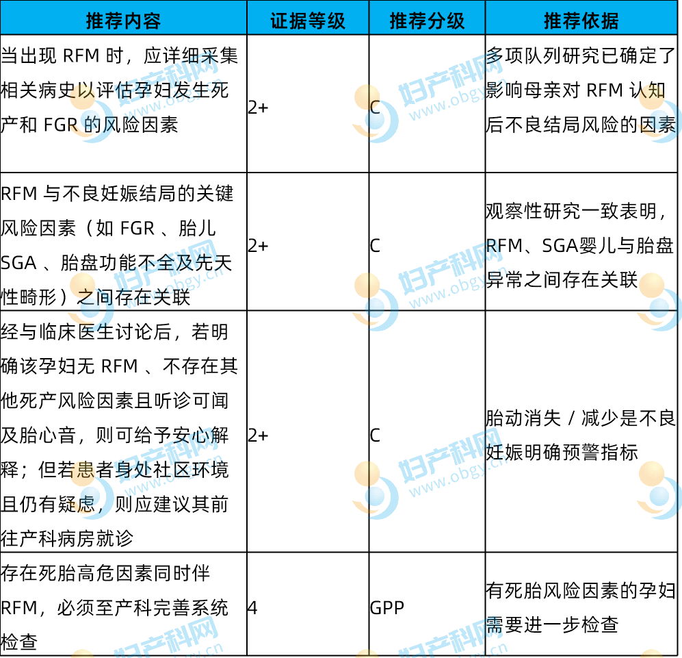
Maternal Risk Factors for Adverse Outcomes Following RFM Presentationa
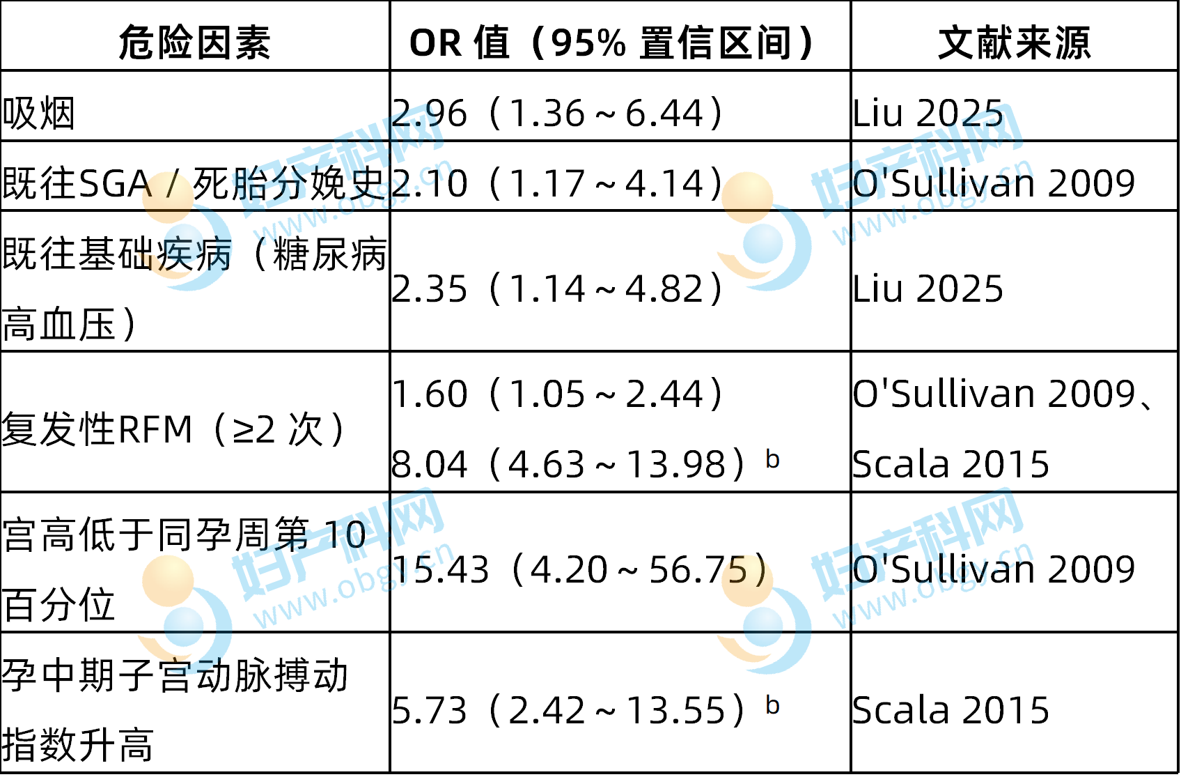
Note a: General population stillbirth risk factors (e.g., nulliparity) are excluded from this table as they demonstrate no independent correlation with adverse outcomes after RFM presentation. Individualized patient assessment remains mandatory for all clinical cases. Note b: Relative risk for delivery of an SGA infant.Additional high-risk factors: Advanced maternal age, obesity, hypertensive disorders of pregnancy, placental abnormalities, prior adverse obstetric history. Maternal smoking and pre-gestational medical disease represent the strongest independent risk factors for adverse outcomes; obesity elevates stillbirth risk (OR=1.8) and FGR risk (OR=1.6).
8.2 Initial Clinical Physical Examination
8.3 Clinical Utility of Cardiotocography

(1) Minimum CTG recording duration is 20 minutes. Reactive tracings with fetal heart rate accelerations accompanying movements confirm intact fetal autonomic function. Persistent absence of accelerations for 80 minutes in term gestations strongly indicates fetal distress.
(2) Clinical cohort data: Among 3,014 Norwegian women presenting with RFM, 3.2% demonstrated abnormal CTG tracings. Women with abnormal tracings carried a 7-fold elevated risk of perinatal adverse outcomes (adjusted OR=7.1); persistent non-reactive tracings carried a stillbirth rate of 26‰ versus 1.9‰ for reactive tracings. 5% of patients with persistent abnormal CTG develop poor neonatal outcomes, while 95% of women with normal tracings experience uncomplicated delivery.
8.4 Standardized Ultrasound Imaging Protocols
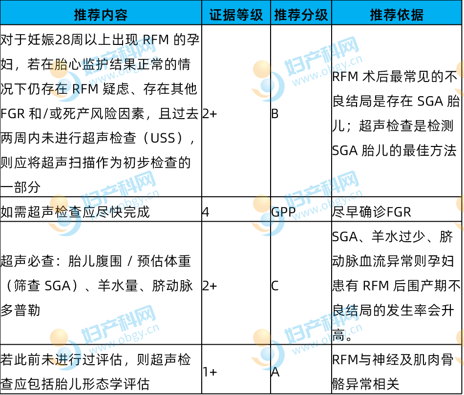
(1) Universal routine ultrasound is not indicated for all women with RFM. The large AFFIRM cluster RCT confirmed mandatory ultrasound for every RFM presentation fails to reduce stillbirth rates and increases rates of labour induction and caesarean delivery.
(2) Indications for targeted ultrasound: Persistent subjective RFM despite normal CTG, presence of clinical high-risk factors, no ultrasound within 14 days, recurrent RFM (≥2 episodes; ultrasound abnormality detection rate rises markedly with recurrent presentation, with one new SGA case identified per 48 ultrasound scans).
(3) Isolated single-episode RFM with no clinical risk factors, normal CTG and normal amniotic fluid volume requires no follow-up ultrasound surveillance.
(4) Isolated abnormal umbilical artery Doppler velocimetry occurs rarely (0.2%–1.4%) yet confers an extremely poor fetal prognosis. Reduced middle cerebral artery (MCA) Doppler velocimetry and low cerebroplacental ratio (CPR) are predominantly observed in women with recurrent RFM. For term RFM ≥37 weeks’ gestation, a CPR <1.1 may serve as a reference threshold for elective early delivery planning.
8.5 Biophysical Profile (BPP)

Current clinical evidence is inadequate to support BPP as a standard screening test for RFM presentations; BPP shall only be utilized as an adjunctive assessment for high-risk pregnancies. A normal BPP carries a false-negative rate of merely 0.07%, with virtually no short-term intrauterine fetal death events documented following normal BPP results. Small-cohort studies indicate BPP cannot reliably stratify fetal prognosis.
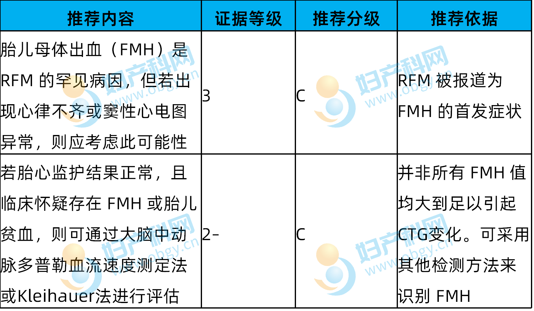
8.6 Clinical Evaluation of Fetomaternal Haemorrhage (FMH)
9. Clinical Management Principles for Abnormal Fetal Growth or Cardiotocography Findings
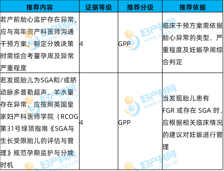
(1) CTG tracings consistent with acute fetal distress trigger immediate evaluation for urgent delivery. Suspicious, equivocal tracings require repeat serial CTG monitoring at increased intervals.
(2) Confirmed SGA, abnormal umbilical artery Doppler flow or oligohydramnios mandates clinical management aligned with RCOG Green-top Guideline No.31 (SGA/FGR guideline), including intensified antenatal surveillance and standardized delivery timing thresholds.
10. Follow-Up Surveillance Protocols for Women with Unremarkable Diagnostic Workup Post RFM Presentation
11. Indications for Induction of Labour Following Normal Diagnostic Testing
12. Management of Recurrent RFM
Definition of recurrent RFM: Two or more episodes of subjective RFM within 21 days after 26 weeks’ gestation.
13. RFM Management in Multiple Gestation Pregnancies
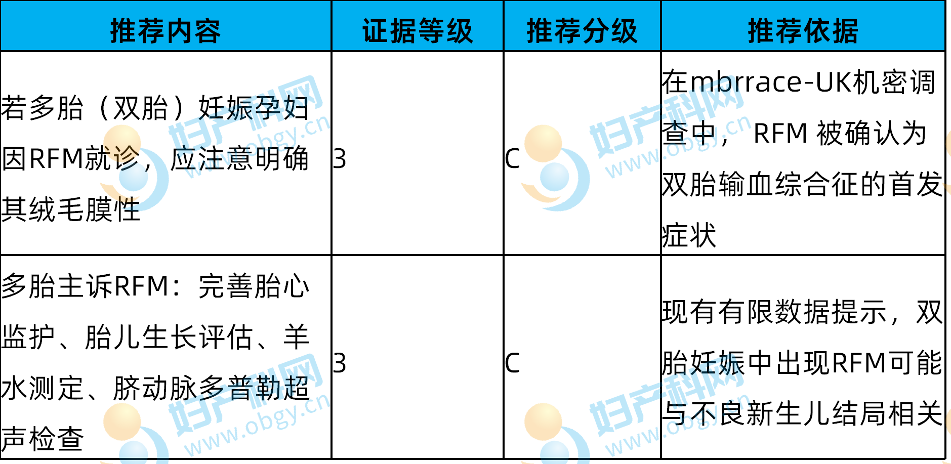
(1) Total fetal movement counts in twin pregnancies are lower than singleton gestations; individual fetuses maintain independent sleep-wake cycles with asynchronous movement patterns.
(2) Isolated subjective RFM in late twin gestation warrants heightened suspicion for twin-twin transfusion syndrome and selective fetal growth restriction. RFM presentation after 34 weeks’ twin gestation carries markedly elevated risks of perinatal death and neonatal intensive care admission.
14. RFM Management Protocols Prior to 28⁺⁰ Weeks’ Gestation
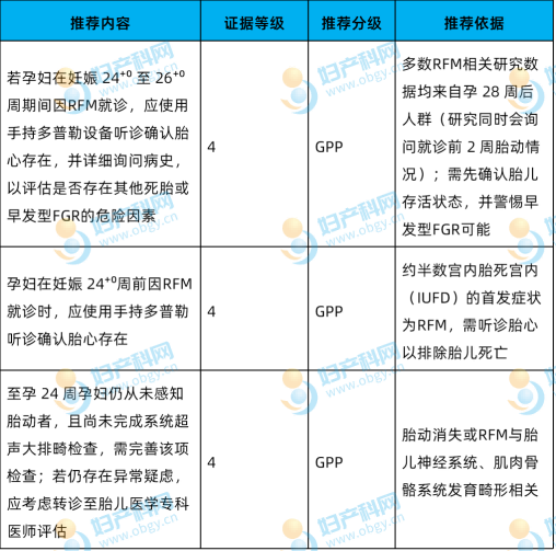
(1) 24⁺⁰–26⁺⁰ weeks’ gestation: Handheld Doppler fetal heart confirmation, comprehensive risk factor history taking to screen for early-onset FGR. Routine CTG is not indicated; targeted ultrasound is only performed if fetal growth restriction is clinically suspected.
(2) <24⁺⁰ weeks’ gestation: Only handheld Doppler auscultation to confirm fetal cardiac activity is required. Women with complete absence of perceived movements by 24 weeks’ gestation undergo full anatomical survey ultrasound, with specialist fetal medicine referral for abnormal findings, as this presentation carries high suspicion of fetal neuromuscular developmental anomalies. Prospective clinical studies investigating RFM before 28 weeks’ gestation remain scarce, with no universal standardized ultrasound or CTG surveillance protocols established.
15. Clinical Management of Abnormally Violent Fetal Movements

Appendix: Standard Clinical Management Flowchart for RFM
Disclaimer:
This document is intended solely for educational reference among medical professionals and does not constitute definitive clinical practice guidance. All clinical decisions shall be individualized based on patient clinical status, institutional resources and latest clinical guidelines; multidisciplinary consultation shall be arranged for complex cases as required.
Article Source:
M.Whitworth, M.Fisher, A.Heazell, Royal College of Obstetricians and Gynaecologists, “Reduced Fetal Movements Green-Top Guideline No. 57,” BJOG: An International Journal of Obstetrics & Gynaecology (2026): 1–23, https://doi.org/10.1111/1471-0528.70198.Editor-in-Charge Reviewer
Editor-in-Charge: Qinghuan
Reviewer: Ma Ye