





















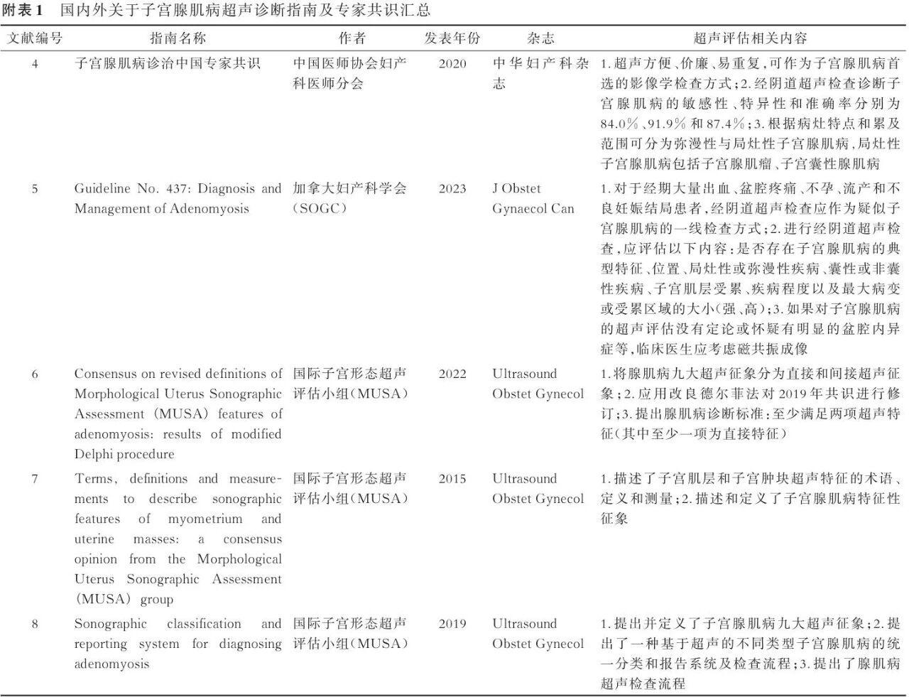
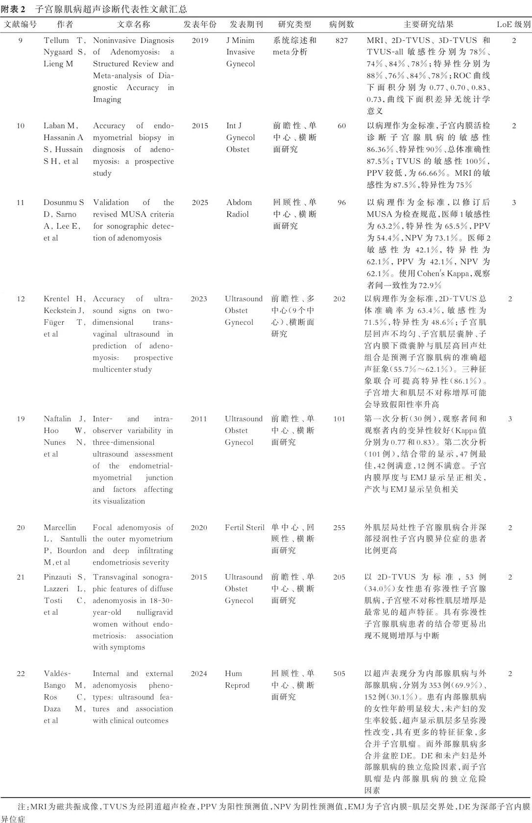
Adenomyosis is a common gynecological disease among women of reproductive age, significantly impairing quality of life and reproductive health. Transvaginal ultrasound is the first-line imaging modality for adenomyosis; however, there is currently a lack of standardized guidance regarding ultrasound examination for this condition in China. In view of this, the Gynecological and Obstetric Ultrasound Group of the Chinese Society of Ultrasound in Medicine, in conjunction with relevant domestic experts, formulated this consensus based on reference to relevant domestic and international guidelines and the latest research literature following multiple rounds of discussion. This consensus covers the standardization and procedures of ultrasound examination for adenomyosis, sonographic manifestations, key points for diagnosis and differential diagnosis, and standardized reporting. It aims to promote the homogenization and standardization of ultrasound examination and assessment for adenomyosis in China, thereby elevating the level of clinical diagnosis and evaluation. Relevant domestic and international guidelines and consensus statements referenced herein are detailed in Appendix Table 1, and representative literature is summarized in Appendix Table 2.
I. Clinical Indications for Ultrasound Screening of Adenomyosis
Ultrasound examination should be performed to confirm or exclude adenomyosis when patients present with symptoms such as dysmenorrhea, pelvic pain, dyspareunia, or menorrhagia; have a history of low fertility or adverse pregnancy outcomes; or are found to have unexplained uterine enlargement upon gynecological examination.
II. Basic Requirements for Ultrasound Examination
1. History Taking: Includes sexual history, clinical symptoms, obstetric history, and other relevant medical history.
2. Examination Route: Transvaginal ultrasound (TVUS) is the preferred method. Transrectal ultrasound (TRUS) is recommended for patients without sexual experience. If imaging quality via TVUS/TRUS is suboptimal, transabdominal ultrasound (TAUS) may be combined.
3. Equipment: It is recommended to use ultrasound equipment equipped with high-resolution endocavitary probes or convex array probes. Where conditions permit, endocavitary probes with three-dimensional (3D) imaging capabilities may be utilized.
4. Preparation:
◦ TVUS/TRUS: Bladder should be emptied prior to examination. No special bowel preparation is usually required for TRUS.
◦ TAUS: The bladder needs to be moderately distended.
III. Normal Uterine Myometrial Anatomy and Sonographic Appearance
The uterus consists of the uterine body (corpus) and cervix, demarcated by the internal os. Arcuate arteries run along the uterine serosa in the outer third of the myometrium, giving off radial arteries that penetrate the muscle layer vertically. On ultrasound, the normal myometrium appears as a homogeneous iso-echoic layer extending from the outer edge of the endometrium to the serosa (Figure 1).
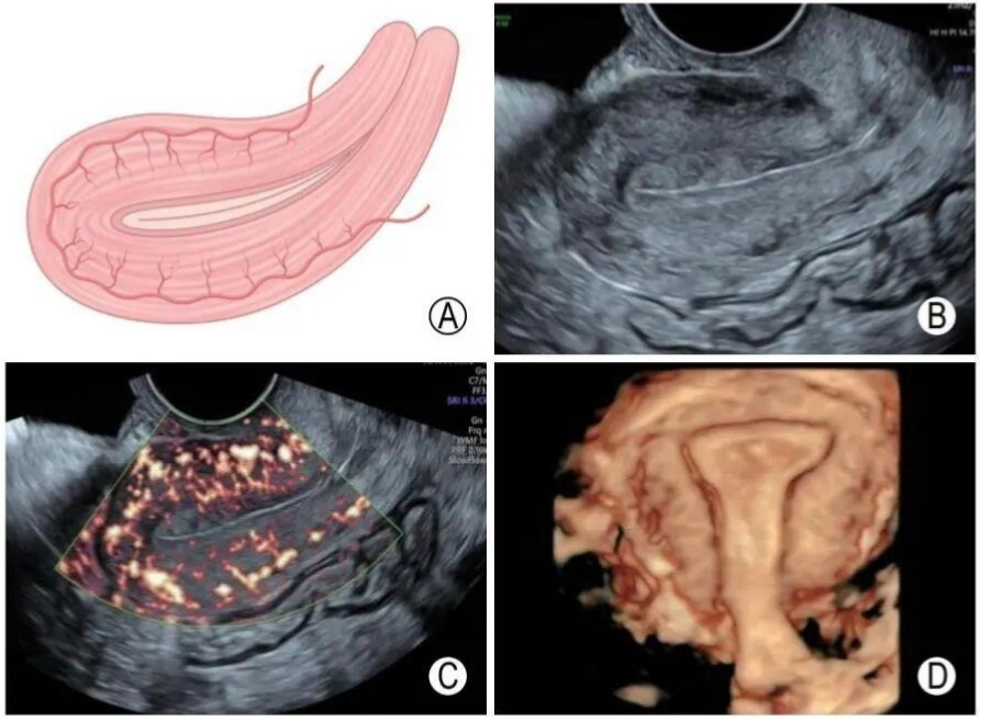
Figure 1. Sonographic appearance of the normal uterine myometrium.
A: Schematic diagram of uterine myometrium and arcuate vessels;
B: 2D grayscale image of the uterine myometrium;
C: Power Doppler image of the uterine myometrium;
D: 3D ultrasound image showing the myometrium and uterine cavity.
➱ The uterine corpus myometrium can be divided into three layers based on echo texture:
1. Junctional Zone (Inner Myometrium/Zone): Composed of longitudinal smooth muscle fibers, located at the interface between the endometrium and myometrium. It appears as a band-like hypoechoic zone parallel to the endometrium on both 2D and 3D ultrasound.
2. Middle Myometrium: Composed of interwoven muscle fibers, situated between the arcuate vessels and the junctional zone, appearing as medium echogenicity.
3. Outer Myometrium (Subserosal Layer): The muscle layer between the serosa and the arcuate vessels, appearing as medium echogenicity.
Uterine Body Measurements: Measure the longitudinal diameter, anteroposterior (AP) diameter, and transverse diameter. The distance from the fundus to the internal os on the longitudinal section is the longitudinal diameter; the maximum diameter perpendicular to the longitudinal axis is the AP diameter. The transverse diameter is measured on the fundal transverse section slightly below the cornua. The normal reference range for the sum of the three diameters in parous women is typically 15–18 cm, and 12–15 cm in nulliparous women. Measurements exceeding these ranges suggest uterine enlargement.
IV. Sonographic Manifestations of Adenomyosis
Common ultrasound signs of adenomyosis include uterine enlargement, asymmetric myometrial thickening, heterogeneous myometrial echotexture, and fan-shaped acoustic shadowing. Other related signs include subendometrial hyperechoic lines or "buds," irregularity or discontinuity of the junctional zone, and translesional blood flow signals (Figure 2).
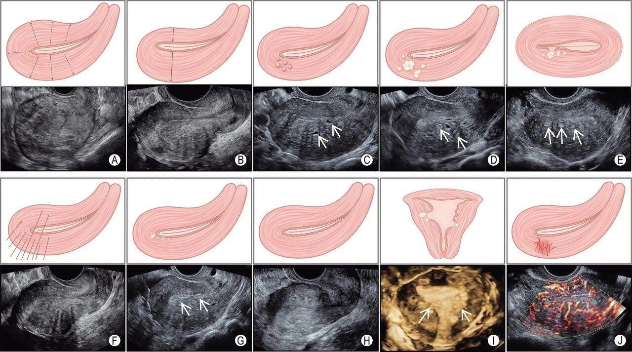
Figure 2. Schematic diagram and ultrasound findings of adenomyosis.
A: Globular uterus; B: Asymmetric myometrial thickening; C: Intramyometrial cystic structures (arrow); D, E: Patchy hyperechoic areas within the myometrium (arrows); F: Fan-shaped acoustic shadowing; G: Subendometrial hyperechoic line and buds; H: Irregular junctional zone; I: 3D ultrasound showing disruption of the junctional zone (arrow); J: Translesional blood flow.
1. Common Ultrasound Signs
(1) Uterine Enlargement: Adenomyosis often manifests as enlargement of the uterine corpus. In severe cases, the uterus may assume a "globular" configuration where the longitudinal, AP, and transverse diameters are nearly equal—a typical sign termed a globular uterus.
(2) Asymmetric Myometrial Thickening: Adenomyosis can cause myometrial thickening. An AP wall thickness difference ≥5 mm allows for the diagnosis of asymmetry. Measurements should be taken on the longitudinal section perpendicular to the endometrium, measuring the maximum thickness of the anterior and posterior walls from the serosa to the outer edge of the endometrium (including the junctional zone). Examinations should avoid the period of uterine contraction to minimize measurement error.
(3) Heterogeneous Myometrial Echoes: This can manifest as intramyometrial cystic structures or focal hyperechoic areas. Cystic structures may appear anechoic, hypoechoic, "ground-glass," or mixed echoic, sometimes surrounded by a hyperechoic rim; Color Doppler should be used to differentiate these from intramyometrial vessels. Focal hyperechoic areas refer to hyperechoic regions within the myometrium unrelated to the endometrium. These findings are direct sonographic signs of ectopic endometrial implants within the myometrium.
(4) Fan-shaped Acoustic Shadowing: A hypoechoic fan-shaped shadow may be visible behind the lesion, alternating with linear hyperechoic lines. This finding is often associated with microcystic/cystic structures within the myometrium.
2. Other Ultrasound Signs
(1) Subendometrial Hyperechoic Line or Buds: Linear hyperechoic lines beneath the endometrium often run perpendicular to the endometrial canal; bud-like hyperechoic foci may be single or multiple. These are direct signs of endometrial invasion into the myometrium through the basal layer.
(2) Irregularity or Disruption of the Junctional Zone: If a regular, continuous band-like hypoechoic zone is visualized in any plane, the junctional zone is considered intact; otherwise, it is deemed irregular and/or discontinuous.
(3) Translesional Blood Flow: Vessels within the myometrium run perpendicular to the endometrial cavity or serosa, traversing the lesion area.
3. Classification of Adenomyosis and Sonographic Findings
Based on the distribution of lesions on sonography, adenomyosis is classified into diffuse, focal, mixed, and special types (Figures 3, 4).
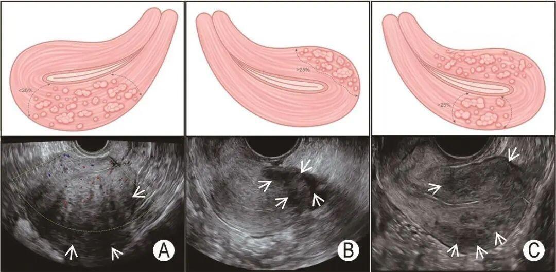
Figure 3. Schematic and ultrasound findings of different types of adenomyosis.
A: Posterior wall diffuse adenomyosis;
B: Posterior wall focal adenomyosis;
C: Mixed-type adenomyosis.
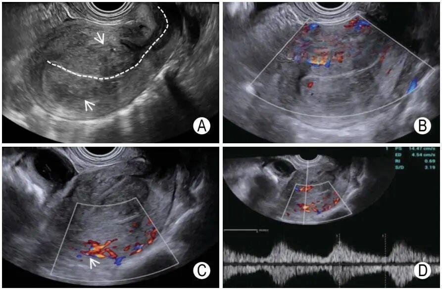
Figure 4. Sonographic features of endometrial polypoid adenomyoma.
A: The lesion appears as an iso-echoic mass with clear boundaries and regular edges, homogeneous internal echoes (white arrow, white dashed line indicates endometrial line);
B, C: Blood supply from the anterior and posterior myometrium is visible, showing a dendritic pattern (white arrows);
D: Moderate resistance arterial spectrum can be measured.
(1) Diffuse Adenomyosis: Diagnosed when the proportion of normal myometrium surrounding the lesion is <1/4, or when multiple focal lesions involve ≥1/4 of the myometrium. This type mostly involves the inner and middle myometrium, exhibiting classic signs such as asymmetric thickening, globular uterus, and junctional zone disruption. Patients usually have significant symptoms, mainly dysmenorrhea, menorrhagia, and irregular vaginal bleeding.
(2) Focal Adenomyosis: Diagnosed when the lesion is surrounded by ≥1/4 of normal uterine myometrium. This type often involves the outer myometrium and is easily complicated by deep infiltrating endometriosis and pelvic adhesions. If the focal lesion has relatively clear borders, it is termed an adenomyoma; if obvious cystic cavities are visible, it is termed cystic adenomyosis. Cystic adenomyosis is more common in young women (<30 years) and is characterized by severe progressive, drug-refractory dysmenorrhea, requiring differentiation from accessory cavitated uterine malformations and cystic degeneration of fibroids.
(3) Mixed-Type Adenomyosis: Diagnosis is made when two types of lesions coexist.
(4) Special Types of Adenomyosis
Endometrial Polypoid Adenomyoma (also known as AdenoFibromatous Polyp) is a rare benign mixed epithelial and mesenchymal polypoid uterine tumor. Histologically, it consists of interwoven uterine smooth muscle fibers, endometrial glands, and endometrial stroma. Based on whether glands show atypia, it is divided into typical and atypical polypoid adenomyomas, accounting for approximately 1.3%–8.0% of endometrial polyps. Its ultrasound appearance is similar to endometrial polyps; due to the presence of smooth muscle components, it often appears iso-echoic or hypoechoic and may show cystic changes. Color Doppler can reveal pedicle vessels with a dendritic blood flow distribution. Ultrasound cannot reliably distinguish between typical and atypical subtypes; diagnosis relies on histopathology.
V. Differential Diagnosis
1. Uterine Fibroids (Leiomyoma): Fibroids typically have clear borders with a peripheral circular blood flow signal. When cystic degeneration occurs, differentiation is difficult. Contrast-enhanced ultrasound (CEUS) or MRI can aid differentiation: fibroids typically show peripheral ring enhancement with contrast filling from the periphery inward, distinguishing them from the translesional blood flow of adenomyosis.
2. Endometrial Cancer with Myometrial Invasion: Common in middle-aged and elderly women, often presenting with abnormal endometrial thickening, irregular shape, and heterogeneous echoes. Color Doppler shows abundant abnormal blood flow. When invasion disrupts the junctional zone, differentiation from adenomyosis is challenging. In adenomyosis, the endometrium may be uniform or heterogeneous, but internal blood flow is relatively sparse.
3. Myometrial Malignancies (e.g., Uterine Sarcoma): Tumors grow rapidly, appear hypoechoic, and may show extensive necrosis (mixed echoes). Posterior acoustic shadowing is rare. Color Doppler shows abundant, tortuous, and chaotic blood flow within the lesion, differing from the penetrating translesional flow of adenomyosis.
VI. Precautions
1. Adenomyosis often coexists with endometriosis. If a patient also has endometriosis, it is recommended to follow relevant expert consensus for standardized ultrasound examination and clinical management.
2. Ultrasound has certain limitations in the assessment of adenomyosis. Diagnostic accuracy is influenced by operator experience, instrument resolution, and individual patient factors.
VII. Recommended Ultrasound Diagnostic Algorithm for Adenomyosis (Figure 5)
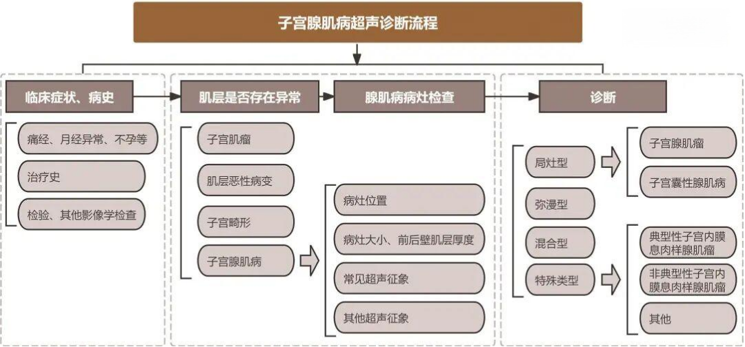
Figure 5. Ultrasound diagnostic algorithm for adenomyosis.
VIII. Reporting
It is recommended to use a structured report format.
[Report Description]
(1) Overall Uterine Assessment
• Uterine Body Size: Longitudinal ___ (mm) × AP ___ (mm) × Transverse ___ (mm)
• Endometrial Thickness: ___ (mm)
• Uterine Morphology: [ ] Normal [ ] Enlarged [ ] Globular Enlargement
• Uterine Contour: [ ] Regular [ ] Irregular
• Endometrial Morphology: [ ] Normal [ ] Displaced/Deformed [ ] Other: _________
• Endometrial Echo: [ ] Homogeneous [ ] Heterogeneous, hyperechoic mass visible, size: ____ mm
(2) Characteristics of Adenomyosis Lesions
• Anterior/Posterior Wall Asymmetry: [ ] Yes, Ant wall: ___ mm, Post wall: ___ mm [ ] No
• Heterogeneous Myometrial Echoes:
◦ (1) Intramyometrial Cystic Structures: [ ] None [ ] Present, size ___ mm;
◦ (2) Focal Hyperechoic Areas in Myometrium: [ ] None [ ] Present;
◦ (3) Fan-shaped Acoustic Shadowing: [ ] None [ ] Present;
◦ (4) Subendometrial Hyperechoic Line/Buds: [ ] None [ ] Present
• Site Involved: [ ] Anterior wall [ ] Posterior wall [ ] Fundus [ ] Other ____
• Junctional Zone: [ ] Regular & Continuous [ ] Disrupted [ ] Irregular Thickening
• Color/Power Doppler: Translesional Blood Flow within Lesion: [ ] Not seen [ ] Visible
(3) Concurrent Lesions
• Concurrent Uterine Fibroids: [ ] None [ ] Present, Number: ___, Largest located at: [ ] Ant wall [ ] Post wall [ ] Fundus, Size: ___ mm;
• Concurrent Ovarian Endometriosis: [ ] None [ ] Present, Location & Size: _________;
• concurrent Pelvic Endometriosis: [ ] None [ ] Present, Location & Size: _________;
[Impression / Recommendations]
[ ] Diffuse type [ ] Focal type [ ] Mixed type [ ] Special type Adenomyosis
[ ] Complicated by Uterine Fibroids
[ ] Complicated by Endometriosis, further examination suggested.
Conclusion
As the primary imaging modality for adenomyosis, ultrasound plays a vital role in screening, diagnosis, and differential diagnosis. It is recommended that sonographers perform standardized examinations of adenomyosis according to the procedures and terminology outlined in this expert consensus. If ultrasound images are atypical or clinical symptoms do not correlate with sonographic findings, the patient's history and other imaging modalities should be integrated for comprehensive assessment.
Source: Gynecological and Obstetric Ultrasound Group, Chinese Society of Ultrasound in Medicine. Expert Consensus on Ultrasound for Adenomyosis (2026 Edition) [J]. Chinese Journal of Ultrasonography, 2026, 35(3): 185-192. DOI: 10.3760/cma.j.cn131148-20260214-00110.
Editor: Lily