





















Obstetrical anal sphincter injury (OASI) is classified as a severe perineal laceration, defined as a partial or complete disruption of the anal sphincter complex, which may extend to the anal mucosa. OASI can be accompanied by significant short-term and long-term sequelae, exerting profound psychological, emotional, economic, and social impacts on women's physical and mental health, family life, and the healthcare system. Among these, long-term complications may appear years or even decades after delivery, severely affecting a woman's quality of life. Therefore, providing timely, professional, and standardized care to OASI patients is crucial for improving their clinical outcomes.
Image Source: JOGC
The Society of Obstetricians and Gynaecologists of Canada (SOGC) Guideline No. 457, "Obstetrical Anal Sphincter Injuries (OASIs) Part I: Prevention, Recognition, and Immediate Management," clearly elucidates the importance of OASI prevention, accurate clinical diagnosis, and adequate primary repair, providing evidence-based guidance for early intervention in OASI.
This guideline (SOGC Clinical Practice Guideline No. 465, "Obstetrical Anal Sphincter Injuries (OASIs) Part II") aims to standardize the postpartum care process for women with OASI and provide scientific, systematic recommendations for long-term management, bridging the early intervention requirements of Guideline No. 457 to form a comprehensive OASI management system.
[Recommendations]
1. Obstetric care providers should refer all postpartum women with a clinical diagnosis of OASI and pregnant women with a history of OASI to a specialized clinic; where no specialized clinic is available, arrange follow-up with an experienced physician and refer to a specialist as needed (Conditional recommendation, Moderate quality evidence).
2. All women with a clinical diagnosis of OASI (Grade 3 or 4) require a birth debriefing, physical and psychological rehabilitation assessment, identification and management of short- and long-term complications, health education, emotional support, counseling regarding mode of delivery for subsequent pregnancies, and guidance on preventing future pelvic floor dysfunction (Strong recommendation, Moderate quality evidence).
3. Women who become pregnant again after OASI require counseling regarding the mode of delivery for the subsequent pregnancy (Strong recommendation, Moderate quality evidence).
4. Trained clinicians should use endoanal ultrasound to assess the structure of the anal sphincter complex and anorectal manometry to assess function, to provide a basis for managing current anorectal symptoms, advising on mode of delivery for subsequent pregnancies, and identifying high-risk women (Strong recommendation, High quality evidence).
5. Women with anorectal symptoms postpartum or women without a clinical diagnosis of OASI during vaginal delivery but who develop symptoms long-term should be referred to an experienced physician for examination and management due to the possibility of missed OASI (Conditional recommendation, Moderate quality, evidence).
6. In institutions without endoanal ultrasound, 2D or 3D transperineal ultrasound may be used as a screening tool for structural assessment after primary repair of the anal sphincter complex; if residual anal sphincter defect is confirmed, refer to a center with endoanal ultrasound for confirmation. Digital rectal examination alone is prohibited for diagnosing residual anal sphincter defects (Strong recommendation, Moderate quality evidence).
7. In institutions without anorectal manometry, clinicians may combine digital rectal examination with endoanal/transperineal ultrasound (2D or 3D) to assess the function of the anal sphincter complex; no validation studies are currently available for this (Conditional recommendation, Low quality evidence).
8. Obstetric care providers should refer all postpartum women with a clinical diagnosis of OASI to pelvic health physiotherapy; if resources are limited, use local or remote resources to initiate treatment as early as 2–6 weeks postpartum (Strong recommendation, High quality evidence).
9. Obstetric care providers should use a collaborative model to provide counseling on mode of delivery for all women with a history of OASI, and document the communication content and the patient's preferred mode of delivery (Strong recommendation, High quality evidence).
10. Obstetric care providers should selectively use episiotomy as needed during subsequent delivery, rather than routine use (Strong recommendation, Moderate quality evidence).
11. For asymptomatic women with no residual anal sphincter defect on postpartum endoanal/transperineal ultrasound and normal anorectal manometry, repeat testing is not required in a subsequent pregnancy, and a trial of vaginal delivery may be offered (Conditional recommendation, Moderate quality evidence).
12. Obstetric care providers should use endoanal ultrasound to assess structure and anorectal manometry to assess the function of the anal sphincter complex; where such equipment is unavailable, 2D/3D transperineal ultrasound and/or digital rectal examination may be selected as alternatives. Both structural integrity and function must be assessed simultaneously to guide counseling and management (Strong recommendation, Moderate quality evidence).
13. In institutions without endoanal ultrasound and anorectal manometry, offer elective cesarean section to women with a previous Grade 4 perineal laceration who do not plan to undergo secondary anal sphincter repair; offer vaginal delivery to women with a previous Grade 4 perineal laceration with moderate-to-severe anorectal symptoms who plan to undergo secondary anal sphincter repair (Conditional recommendation, Moderate quality evidence).
14. Obstetric care providers should recommend elective cesarean section for women with a previous secondary anal sphincter repair (Strong recommendation, Moderate quality evidence).
15. When deciding indications for cesarean section in a subsequent delivery, obstetric care providers should incorporate individualized risk factors for OASI recurrence (Strong recommendation, High quality evidence).
16. Obstetric care providers should document the communication content and the patient's choice regarding mode of delivery after OASI (Strong recommendation, High quality evidence).
I. The Role of Specialized Clinics
Given the potential sequelae of OASI, the optimal approach for postpartum women with a clinical diagnosis of OASI and women pregnant again after OASI is follow-up by a physician experienced in OASI diagnosis and treatment at a specialized clinic. Where referral to a specialized clinic is not possible, follow-up is conducted by an obstetrician, with referral for appropriate investigations and specialized care as needed.
Appropriate specialized resources and investigations can also be accessed in institutions without specialized clinics. While Canada has successfully established several specialized clinics, their development remains incomplete, and many women (especially in remote areas) cannot access specialized care. In the future, multidisciplinary specialized clinics need to be established and improved to help women with OASI nationwide receive professional care and long-term management.
A multidisciplinary specialized team ensures consistency in counseling. The optimal team configuration covers Obstetrics, Urogynecology, Pelvic Health Physiotherapy, Urology, Nursing, Incontinence Nurse Specialists, Colorectal Surgery, Psychology, Psychiatry, Sex Therapy, Social Work, Lactation Consulting, Pharmacy, and Clinical Nutrition.
Ensuring equitable access to specialized services is critical; women should not be unable to obtain the care needed for rehabilitation, long-term health, and informed decision-making for subsequent deliveries due to financial or geographical factors.
➱ Care should focus on the following content:
• Patient birth debriefing, communicating OASI-related delivery circumstances.
* Postpartum rehabilitation assessment, including anorectal and urinary symptoms, wound infection, pelvic organ prolapse, sexual function, pain, emotional and psychological state, and impact on quality of life.
* Providing consistent multidisciplinary health education and support for women and caregivers.
• Early identification and management of short- and long-term OASI-related complications, providing specialized surgical intervention if necessary (e.g., secondary anal sphincter repair, sacral neuromodulation, rectovaginal fistula repair).
* Providing management strategies to reduce the risk of OASI recurrence during subsequent delivery.
* Providing counseling for women to assist them in making an informed choice regarding mode of delivery for subsequent pregnancies.
* Guiding the prevention of future pelvic floor dysfunction.
Delivery related to OASI may be traumatic for women. Care must follow the principles of Trauma- and Violence-Informed Care (TVIC), including creating an emotionally safe clinical environment, acknowledging the potential impact of past trauma or abuse, and adjusting clinical procedures (including physical examinations and counseling) to reduce distress and enhance safety and a sense of control.
Women with OASI may experience psychological sequelae such as anxiety, depression, body image changes, shame, coping difficulties, and social isolation, and may be reluctant to seek medical care, thereby affecting mother-infant relationships, infant development, and family intimacy. Female partners may also experience anxiety and social isolation, suggesting the need to support both the woman and her family.
II. Postpartum and Subsequent Pregnancy Assessment
Assessment of women with a clinical diagnosis of OASI includes history taking, physical examination, and auxiliary investigations. The timing of postpartum assessment varies among specialized clinics; some assess at fixed times, while others assess based on symptom progression. Considering the incidence of early wound complications, initiating follow-up at 1–2 weeks postpartum is reasonable. Postpartum re-evaluation at 6 weeks and subsequent follow-up should screen for OASI-related complications.
Women pregnant again after OASI require counseling regarding mode of delivery for the subsequent pregnancy. Currently, there is no evidence on the optimal assessment time during subsequent pregnancy; factors such as the risk of spontaneous miscarriage, possible pregnancy termination, the cycle of specialized consultation and investigation, the woman's decision-making time, and scheduling of cesarean section need to be comprehensively considered. Expert consensus suggests the optimal assessment time is late pregnancy (after 28 weeks), adjustable based on individual circumstances.
History and Physical Examination
When using digital rectal examination (DRE) alone to assess the function of the anal sphincter complex, the Digital Rectal Examination Scoring System (DRESS) can be used to record resting pressure and squeeze pressure.
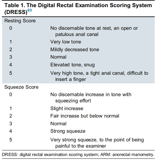
Table 1: Digital Rectal Examination Scoring System (DRESS)
III. Auxiliary Investigations
➱ Purposes of anorectal investigation in women with OASI:
1. Confirm accuracy of clinical diagnosis.
2. Assess structure and function of the anal sphincter complex.
3. Guide management of anorectal symptoms (including surgical planning).
4. Formulate individualized advice on mode of delivery for subsequent pregnancies.
5. Stratify and assess the risk of future anorectal symptoms.
For women with OASI, endoanal ultrasound (EAUS) and anorectal manometry (ARM) should be used to simultaneously assess the integrity and function of the anal sphincter complex; in institutions without relevant equipment, available alternative investigations may be selected.
Endoanal Ultrasound (EAUS)
Endoanal ultrasound is the gold standard for structural assessment of the anal sphincter complex, usually performed after 6 weeks postpartum; early puerperal examination correlates poorly with long-term fecal incontinence.
➱ The probe is inserted into the anal canal to obtain 360° images at 4 levels:
1. Puborectalis muscle;
2. Deep (proximal) external anal sphincter and internal anal sphincter;
3. Superficial (middle) external anal sphincter and internal anal sphincter;
4. Subcutaneous (distal) external anal sphincter.
Determination of Anal Sphincter Complex Integrity
1. Intact: Normal continuity of the anal sphincter complex (no evidence of prior OASI).
2. Scar: Partial or full-thickness disruption of normal continuity of the anal sphincter complex, width <1 hour (<30°), a common finding at the site of primary OASI repair.
3. Defect: Partial or full-thickness disruption of normal continuity of the anal sphincter complex (external and/or internal anal sphincter), width ≥1 hour (≥30°), involving at least 2 of the 3 levels. Defect size is described in clock hours or degrees (12 hours = 360°, 1 hour = 30°).

Figure 1: Endoanal ultrasound images
Inadequacy of OASI Grading
A 2023 retrospective observational study including 615 women referred to tertiary care clinics with a clinical diagnosis of Grade 3a or 3b perineal lacerations showed that 1/11 women had internal anal sphincter injury confirmed by endoanal ultrasound, suggesting a laceration of at least Grade 3c. 11.36% of women with a clinical diagnosis of Grade 3a or 3b perineal laceration showed more severe injury on endoanal ultrasound. Women with under-graded OASI had more significant anorectal symptoms, possibly due to inadequate repair, residual anal sphincter defects, and more severe laceration.
Missed OASI
Women with under-graded OASI receive primary anal sphincter repair, whereas women with missed OASI often miss the opportunity for repair, which can lead to serious long-term adverse consequences. The rate of missed OASI in vaginal delivery is 11.5%–14%, and the miss rate in women sutured for Grade 1 or 2 perineal lacerations is 3%. At 2 months postpartum, 71.5% of women with missed OASI developed moderate anal incontinence. A 2017 retrospective study comparing 32 women with missed OASI and 62 women with primary repaired OASI (1:2 matched) showed that the missed group had more severe anorectal and urinary symptoms at 9.31±6.79 months postpartum. Therefore, women without a clinical diagnosis of OASI but who develop anorectal symptoms postpartum need referral to an experienced physician for examination and management; some women with missed OASI may have no obvious symptoms, but their risk of OASI recurrence in subsequent deliveries and anal incontinence in later life is increased.
Overdiagnosis of OASI
Overdiagnosis refers to a clinical diagnosis of OASI, but endoanal ultrasound shows the anal sphincter complex is intact. The reported overdiagnosis rate is 7%–37%; a Canadian retrospective study of 330 women showed an OASI overdiagnosis rate of 36.8%. Such women can be excluded from the OASI diagnosis and do not need elective cesarean section solely because of OASI.
In summary, diagnostic errors in perineal injury severity suggest the need to strengthen physician training, accurate clinical diagnosis, adequate primary OASI repair, clinical practice audits, and accessibility to appropriate investigations and specialized resources.
Residual Anal Sphincter Defect
The incidence of residual anal sphincter defect after primary OASI repair is 34%–91%, theoretically related to factors such as inadequate primary repair and wound infection/dehiscence. Studies confirm that residual anal sphincter defect is closely related to the occurrence of anorectal symptoms. A large systematic review of 16,111 women with OASI showed that 55% had residual anal sphincter defects, of whom 38% had fecal incontinence; women with defects in both the external and internal anal sphincters had the highest risk of anorectal symptoms. A 2023 Canadian study indicated a weak positive correlation between the size of the residual anal sphincter defect and the severity of anorectal symptoms. Note that some women with residual anal sphincter defects may have no obvious clinical symptoms, but the risk of developing anorectal symptoms in the future is significantly increased in such populations. Therefore, endoanal ultrasound is a key investigative tool for assessing the structural integrity of the anal sphincter complex.
In Institutions Without Endoanal Ultrasound
The accuracy of using digital rectal examination alone to diagnose residual anal sphincter defects is limited, with sensitivity of 12%–82% and specificity of 32%–55%; therefore, endoanal ultrasound remains the gold standard for structural assessment of residual anal sphincter defects, and digital rectal examination alone is prohibited for diagnosing residual anal sphincter defects. If the institution does not have endoanal ultrasound equipment, 2D/3D transperineal ultrasound may be used as a screening tool for residual anal sphincter defects; those with abnormal screening results need referral to a center equipped with endo,anal ultrasound for further confirmation.
3D Transperineal Ultrasound (3D-TPUS)
Definition of residual anal sphincter defect: Partial or full-thickness defect appearing in ≥3 of ≥7 slices for the external anal sphincter, and in ≥2 of ≥5 slices for the internal anal sphincter, width ≥30°, with a sensitivity of 70% and specificity of 69% for diagnosing external anal sphincter defects. Note that this definition still needs re-evaluation, as the measurement consistency between 3D transperineal ultrasound and endoanal ultrasound is poor, with a standard error of measurement of ±16.5° for the external anal sphincter and ±26.4° for the internal anal sphincter. If the same 30° cutoff is used, it may lead to misdiagnosis of 9%–36% of external anal sphincter defects and 4%–15% of internal anal sphincter defects; therefore, further research is needed to determine the optimal critical angle for 3D transperineal ultrasound to diagnose significant defects.
3D transperineal ultrasound has a high negative predictive value for detecting residual anal sphincter defects, 85% for the external anal sphincter and 93% for the internal anal sphincter; for patients with no defect on 3D transperineal ultrasound, no referral for further endoanal ultrasound is needed; its positive predictive value is low, 51% for the external anal sphincter and 37% for the internal anal sphincter; for those with abnormal findings, referral to a center with the relevant equipment is needed for further confirmation.
2D Transperineal Ultrasound (2D-TPUS)
If the institution lacks both endoanal ultrasound and 3D transperineal ultrasound equipment, 2D transperineal ultrasound (i.e., vulvar ultrasound) can be used as a screening tool for residual external anal sphincter defects in women with a clinical diagnosis of OASI. This technique has the advantages of high accessibility, low cost, repeatability, and non-invasiveness; the definition of residual anal sphincter defect is consistent with that of 3D transperineal ultrasound; this test has a high negative predictive value, 86% for the external anal sphincter and 93% for the internal anal sphincter, and those with no defect on examination do not need referral for endoanal ultrasound; its positive predictive value is low, 50% for the external anal sphincter and 63% for the internal anal sphincter, and those with abnormal findings need referral to a center with relevant equipment for further confirmation.
Magnetic Resonance Imaging (MRI)
Literature on magnetic resonance imaging (MRI) as an alternative to endoanal ultrasound for evaluating residual external anal sphincter defects is relatively limited. A small prospective study of 40 women showed moderate inter-rater agreement between 1.5T external phased-array MRI and endoanal ultrasound; another small prospective study of 24 women showed that 3T external phased-array MRI results were comparable to endoanal ultrasound. Currently, larger sample, high-quality studies are still needed to clarify whether magnetic resonance imaging can be used as an alternative diagnostic method in institutions without endoanal ultrasound.
Anorectal Manometry (ARM)
Anorectal manometry can objectively measure the function of the anal sphincter complex; normal parameters vary depending on the equipment.
➱ Key parameters in women with OASI:
• Maximum Resting Pressure (MRP): Maximum pressure in the anal canal at rest, 50%–85% from the internal anal sphincter, 25%–30% from sustained tonic contraction of the external anal sphincter, 15% from the anal cushions.
* Maximum Squeeze Pressure (MSP): Maximum pressure in the anal canal during active contraction and coughing, from voluntary contraction of the external anal sphincter.
* Maximum Increment Squeeze Pressure (iMSP): MSP - MRP, reflecting external anal sphincter function.
Anal sphincter dysfunction can occur after both vaginal delivery and cesarean section. A prospective cohort study of 283 primiparous women showed that regardless of whether OASI was present, anal pressure decreased from late pregnancy to 3 months after vaginal delivery. Women with OASI who have anorectal symptoms and residual sphincter defects are more likely to have impaired anal sphincter function; however, a large retrospective cohort study of 13,548 women with OASI showed that 30.6% of women with no residual defect on endoanal ultrasound and no clinical symptoms still had abnormal anorectal manometry results. The occurrence of anorectal symptoms is multifactorial, and related functional abnormalities may be related to injury to the anal sphincter complex, pudendal neuropathy, connective tissue injury, pelvic floor muscle weakness, etc.
Anal sphincter function may gradually recover over time. For women with abnormal postpartum anorectal manometry results, it is recommended to repeat anorectal manometry in a subsequent pregnancy; the extent of impaired anal sphincter function will affect clinical counseling and decision-making on the mode of delivery for subsequent pregnancies.
In Institutions Without Anorectal Manometry
When no manometry equipment is available, function can be assessed by combining digital rectal examination with endoanal/transperineal ultrasound; no validation studies are currently available. Digital rectal examination is minimally invasive, economical, quick, and easy to perform, assessing resting anal tone, active contraction and sustained contraction (normal ≥5 seconds) of the anal sphincter complex and levator ani muscle, and involuntary contraction during coughing/Valsal,va maneuver.
In patients with fecal incontinence, the consistency between digital rectal examination and manometry is poor for resting pressure and moderate for squeeze pressure; the Digital Rectal Examination Scoring System shows a strong positive correlation with resting and squeeze pressures assessed by traditional manometry, and assessment accuracy is positively correlated with physician experience.
Anorectal manometry remains the optimal means for functional assessment; if temporarily undetectable, it can be completed before a subsequent pregnancy or fecal incontinence surgery.
IV. The Role of Vaginal Estrogen
Postpartum women are prone to hypoestrogen-related vulvovaginal symptoms, mainly due to the hypoestrogenic state caused by anovulation postpartum, which is further aggravated during lactation. The only double-blind randomized controlled trial evaluating the efficacy and safety of postpartum vaginal estrogen included 59 women with Grade 2 or higher perineal lacerations (7 of whom were women with OASI), randomly assigned to a vaginal estrogen group and a placebo group; the vaginal estrogen group used a regimen of 1g, twice a week, for 12 weeks. The results showed: no adverse events occurred in the estrogen group, and there was no significant difference in infant feeding methods between the two groups; at 12 weeks of intervention, the Vulvar Assessment Scale (VuAS) scores improved in both groups, but the improvement was not statistically or clinically significant; the estrogen group had higher subjective satisfaction, but due to insufficient sample size, this difference did not reach statistical significance. This study had obvious limitations: small sample size, low rate of exclusive breastfeeding (<10%), and undefined start time for vaginal estrogen, all of which affect the generalizability of the results. In summary, the role of postpartum vaginal estrogen in improving related symptoms still needs to be confirmed by further high-quality research.
V. The Role of Pelvic Health Physiotherapy
Multiple studies confirm that postpartum pelvic health physiotherapy, especially individualized pelvic floor muscle training (PFMT), has clear overall clinical benefits. A systematic review and meta-analysis of 15 randomized controlled trials showed: compared with expectant management, pelvic floor muscle training significantly improved postpartum pelvic floor dysfunction-related issues, including urinary incontinence, anal incontinence, pelvic organ prolapse, and sexual dysfunction. Subgroup analysis further indicated that pelvic floor muscle training can effectively improve anorectal-related symptoms in women with OASI. OASI is an independent risk factor for persistent stress urinary incontinence and anal incontinence postpartum; individualized pelvic floor muscle training guided by a physiotherapist can effectively correct the above pelvic floor abnormal symptoms. Women with OASI are more prone to multiple pelvic floor dysfunctions within 6 months postpartum, and implementing individualized pelvic floor rehabilitation has definite clinical benefits. Current latest research also believes that early screening and identification, standardized pelvic floor physiotherapy, and whole-process rehabilitation management for women with difficult labor can effectively optimize the quality of postpartum clinical care for this population.
A clinical study included 50 women with OASI, randomly assigned to a standard care group and a pelvic health physiotherapy group; the latter's intervention included biofeedback-assisted pelvic floor muscle training, core deep muscle group exercises, and rehabilitative manual therapy; initiated at 6 weeks postpartum, efficacy was assessed at 12 weeks postpartum. Results showed: there was no significant difference between the two groups in fecal incontinence quality of life scores and anorectal manometry-related indicators, but improvements were seen in both groups in the dimensions of coping ability and depressive mood. This study had obvious limitations: unbalanced baseline data between groups, higher proportion of Grade 4 perineal lacerations in the intervention group; baseline clinical symptoms of the enrolled population were mild; the follow-up observation period was only 12 weeks; and the intervention course and frequency were insufficient, as the internationally recognized effective intervention regimen is standardized intervention for 12 weeks, 3–4 times a week.
A 2021 randomized controlled trial included 50 women with OASI, randomly assigned to a standard care group and a standard care plus pelvic physiotherapy group; pelvic physiotherapy was initiated at 2 weeks postpartum using an individualized multi-dimensional rehabilitation model covering health education, manual therapy, therapeutic exercise, neuromuscular retraining, and physical agent therapy. Follow-up at 12 weeks postpartum showed: pelvic floor function, urinary and anorectal-related symptoms improved significantly in both groups compared to 2 weeks postpartum; however, the pelvic physiotherapy group showed more prominent improvements in pelvic floor symptoms and somatic distress, with a more significant decrease in Pelvic Floor Distress Inventory (PFDI-20) scores.
Pelvic health physiotherapy can effectively improve clinical symptoms in women with anal incontinence after OASI. Clinical data show that about 30% of women with OASI have fecal incontinence, and pelvic floor muscle training can effectively alleviate such symptoms. A clinical study included 60 women with fecal incontinence at 12 weeks postpartum, randomly assigned to two groups both using biofeedback pelvic floor training; results showed that 96% of the enrolled women had residual external anal sphincter defects suggested by ultrasound, and both groups reported significant improvements in anal continence function scores and anorectal manometry-related indicators. Another randomized controlled trial included women with anal incontinence at 1 year postpartum (including those with and without OASI), randomly assigned to a written information group and a physiotherapy group; the physiotherapy group used a 6-month individualized pelvic floor muscle training program, with 4–6 visits. Results showed that the physiotherapy group had a significant reduction in St. Mark's incontinence score, and symptoms such as fecal urgency and gas incontinence were significantly improved.
A recent qualitative study also supports the clinical benefits of pelvic physiotherapy: interviews with 14 women with OASI found that pelvic physiotherapy gave them a subjective experience of "empowerment" and "reassurance," further confirming the clinical value of this treatment modality.
Prevention of Perineal Injury
Current research shows that routine antenatal pelvic floor muscle training has not been proven to reduce the risk of perineal injury. However, a meta-analysis on OASI (including 30 studies with 6,691 subjects) showed that antenatal pelvic floor muscle training improved maternal and neonatal outcomes and had a preventive effect on urinary incontinence and OASI; this conclusion supports the inclusion of antenatal pelvic floor muscle training into routine antenatal care and health training systems.
Start Time and Mode of Pelvic Health Physiotherapy
For women with OASI, the initiation of pelvic health physiotherapy needs to balance perineal tissue healing and pelvic floor muscle repair, and it is recommended to start as early as possible postpartum. Existing evidence supports that this treatment can be implemented starting at 2–6 weeks postpartum. In terms of treatment mode, face-to-face treatment is the gold standard for pelvic health physiotherapy; if face-to-face treatment is not available, telemedicine and mobile health applications based on evidence-based medicine have definite efficacy. If medical resources are limited, alternative training modes such as yoga and Pilates can be selected, combined with online rehabilitation resources to carry out pelvic floor rehabilitation training.
VI. Mode of Delivery for Subsequent Pregnancy
Among women with a history of OASI, 60%–75% will become pregnant again, and 49% of women experience anxiety during the subsequent pregnancy.
Counseling on Mode of Delivery for Subsequent Pregnancy
All women with a history of OASI need to receive counseling on mode of delivery for subsequent pregnancy and shared decision-making guidance through a multidisciplinary collaboration model, while standardizing the recording of doctor-patient communication content and the patient's preference for mode of delivery.
➱ When counseling women with a history of OASI on the mode of delivery for a subsequent pregnancy, the following factors should be comprehensively considered:
1. Current physical and psychological sequelae after OASI surgery and their impact on quality of life;
2. Physical examination and related auxiliary investigation results;
3. Future fertility desires and plans;
4. Benefits and potential risks of vaginal delivery and cesarean section;
5. Risk of OASI recurrence (rOASI), including individualized risk factors and protective factors;
6. Risk of occurrence or worsening of anorectal symptoms under different modes of delivery;
7. History of previous pelvic floor surgery or sacral neuromodulation (SNM) treatment;
8. Personal preferences of the patient and family regarding mode of delivery.
The main concerns of women with a history of OASI regarding subsequent delivery focus on OASI recurrence and the occurrence or worsening of anorectal symptoms. Clinical data show that 75% to 95% of women are willing to follow physician recommendations based on endoanal ultrasound and anorectal manometry results, fully reflecting the core position of shared decision-making in the choice of delivery mode; another 34.9% of women indicated that the counseling process directly affected their final decision.
Risk of Anorectal Symptoms After Subsequent Delivery
The impact of subsequent delivery on anorectal symptoms currently has no unified evidence-based conclusion; some studies suggest it can cause new onset or worsening of symptoms, while others show no significant difference. Even slight anorectal symptoms can significantly affect a woman's quality of life.
A systematic review and meta-analysis of 27 studies showed that subsequent delivery did not significantly worsen anorectal symptoms, and there was no significant difference in the occurrence of anorectal symptoms after vaginal delivery and cesarean section; however, this conclusion is constrained by factors such as sample size, study quality, and heterogeneity, and the level of evidence is limited.
Currently, there is only one randomized controlled trial, including 222 women with sphincter defects confirmed by endoanal ultrasound and no obvious clinical symptoms, comparing the long-term prognosis of elective cesarean section and vaginal delivery; results showed no statistical difference in the incidence of fecal incontinence at 6–8 weeks and 6 months postpartum. This study had obvious limitations: the definition standard for significant sphincter defect is not yet uniform; only 14.7% of the enrolled population had Grade 3 perineal lacerations, and Grade 4 laceration cases were excluded; the mode of first delivery was forceps-assisted in as high as 91.0%, while vacuum-assisted delivery populations were excluded; routine anorectal manometry to assess pelvic floor function was not performed, the follow-up period was short, and subgroup stratified analysis by residual defect size was not performed.
Another systematic review and meta-analysis of 14 studies involving 977 women pointed out that due to insufficient sample size, uneven methodological quality, and high heterogeneity of existing studies, the optimal mode of delivery for women with a history of OASI cannot be clarified yet. A 2024 systematic review and meta-analysis covering 9 studies also believes that the current quality of evidence is low, and it cannot be confirmed that elective cesarean section can effectively prevent secondary anal incontinence in women with a history of OASI; further high-quality evidence-based research in this field is still needed.
Long-term Anorectal-Related Symptoms After Subsequent Delivery
The incidence of long-term fecal incontinence after subsequent delivery in women with OASI can reach 12%–54%; 41.0% after Grade 3 perineal anal sphincter injury and as high as 58.8% after Grade 4 injury. Those who had OASI during first delivery and experienced transient or permanent anal incontinence have an increased risk of long-term fecal incontinence, and this prognosis is not significantly associated with the mode of subsequent delivery.
The incidence of anorectal-related symptoms is highest in the puerperium, which can be alleviated as postpartum pelvic floor function gradually recovers, and will increase again in old age due to physiological decline of anal sphincter function. Long-term sequelae caused by OASI can appear years or decades after the initial injury, seriously affecting the long-term quality of life of women. Currently, no special study compares the differences in the impact of elective cesarean section and vaginal delivery on long-term anorectal symptoms in women with a history of OASI.
Women without a history of OASI and women with good healing after primary OASI repair may also experience anorectal discomfort symptoms, and their onset is caused by the combined action of multiple factors, mainly including residual sphincter defects, decreased pelvic floor muscle strength, and pudendal nerve injury and lesions. Pelvic floor muscle contraction function is positively correlated with proximal anal canal pressure; the higher the pelvic floor muscle strength level, the milder the anorectal-related symptoms. Clinical data show that about 10% of women with detected residual sphincter defects at 9 weeks postpartum and no conscious symptoms may develop new-onset fecal incontinence after 3 years; some initially asymptomatic women may also develop related symptoms in old age, especially after menopause.
The external anal sphincter, internal anal sphincter smooth muscle, and anal canal connective tissue all have estrogen receptors. Pelvic floor function declines physiologically with age, which can induce or aggravate anorectal symptoms. In women with a history of OASI aged 52 and older, the incidence of fecal incontinence shows a clear upward trend; at the age of 60, the incidence of fecal incontinence in women without a history of OASI, with a single history of OASI, and with two histories of OASI is 19.8%, 36.8%, and 48.8%, respectively.
At this stage, high-quality clinical research still needs to be carried out to further explore the long-term impact of subsequent pregnancy and delivery on pelvic floor function and women's quality of life, and to compare and analyze the incidence and severity of anorectal symptoms in the stages of simple OASI postpartum, after subsequent delivery, and long-term follow-up, to provide an evidence-based basis for the formulation of clinical intervention strategies.
Risk of OASI Recurrence (rOASI)
Women with a history of OASI have a significantly higher risk of OASI recurrence during subsequent delivery than multiparous women without a history of OASI. According to literature reports, the OASI recurrence rate is 2.0%–13.4%, while the OASI incidence in women without a history of OASI is only 1.5%. A 2014 study in Ontario, Canada, including 1,923 women delivering again after OASI showed that their risk of OASI recurrence was 5.3%. Clinically, most women with a history of OASI will not have a recurrence during subsequent delivery; the most common type of laceration during subsequent delivery is Grade 2 (incidence 59.4%). Notably, women with a history of OASI who had a subsequent vaginal delivery without recurrence still have an increasing trend in the risk of OASI recurrence (recurrence rate 3.1%) when they subsequently deliver vaginally again.
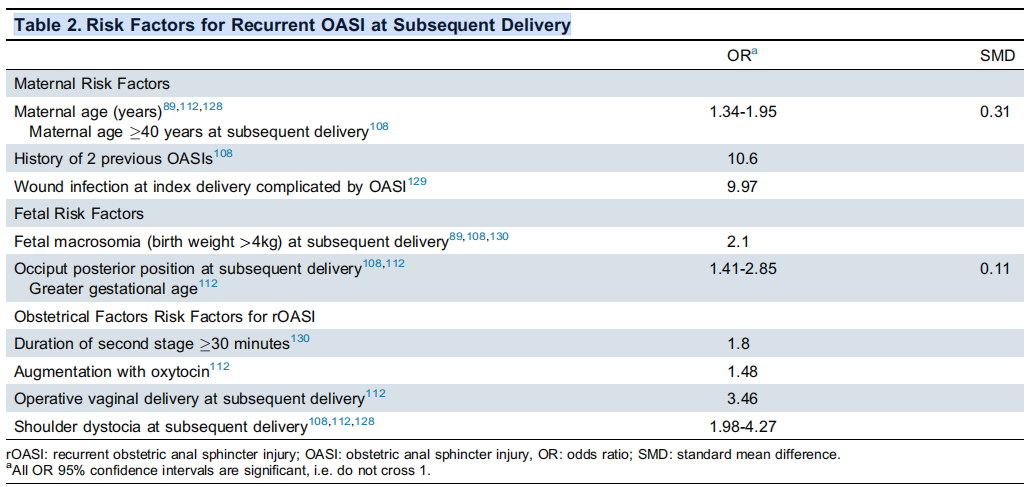
Table 2: Risk factors for recurrent OASI
During clinical counseling, the relevant risk factors for OASI recurrence must be clearly informed to the parturient, most of which are unchangeable (such as maternal age) or only appear during labor (such as oxytocin for uterine contraction, midwifery operations, shoulder dystocia, etc.). In addition, about 0.3% of women with OASI will have a traumatic cloacal defect, mostly caused by dehiscence of a Grade 4 laceration wound; such women need to be highly alert to the risk of wound and related tissue dehiscence when undergoing vaginal delivery again.
Regarding protective factors for OASI recurrence, a systematic review and meta-analysis included 11 clinical studies aimed at analyzing the impact of episiotomy during subsequent delivery on the risk of OASI recurrence; no association was found between the two, suggesting that episiotomy is neither a risk factor nor a protective factor for OASI recurrence. This study had certain limitations, i.e., it did not distinguish the specific angle of episiotomy (median/lateral). Recent special research on episiotomy shows that performing episiotomy during subsequent delivery can reduce the risk of OASI recurrence, and the number of preventive episiotomies required is small; therefore, episiotomy can be considered to prevent OASI recurrence if there are clinical indications.
In summary, the effectiveness of various measures to prevent OASI recurrence during labor still needs further clinical research to be verified. In clinical work, strengthening health consultation for parturients, promoting doctor-patient shared decision-making, and performing selective episiotomy according to clinical situations are important for preventing OASI recurrence.
Management of Recurrent OASI
For women with a history of OASI who choose vaginal delivery, careful examination is required after delivery referring to Guideline No. 457 to clearly identify whether there is OASI recurrence; once confirmed, wound repair and postpartum care should be performed according to the relevant recommendations of this guideline.
Women with two histories of OASI have a significantly increased risk of OASI recurrence (OR=10.6) when undergoing vaginal delivery again, and women with recurrent OASI have more serious anorectal-related symptoms. An observational study compared the long-term prognosis of women without a history of OASI, with 1 history of OASI, and with 2 histories of OASI; results showed that 20 years after the first delivery, the severity of anal incontinence and its impact on life in women with 2 histories of OASI were significantly higher than those in women with 1 history of OASI; no significant difference was found between the two groups in long-term lower urinary tract symptoms and other pelvic floor dysfunctions.
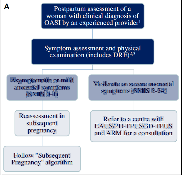
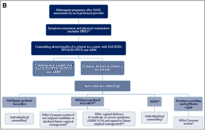
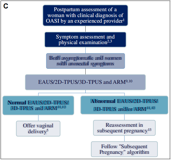
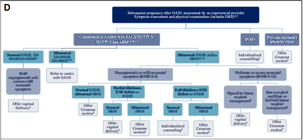
Figure 2: Postpartum management and subsequent pregnancy flowchart (A, B, C, D)
Instructions Related to the Counseling Flowchart
The optimal mode of delivery for women with a history of OASI who are pregnant again currently requires further clinical research for clarification. Recommendations regarding the mode of delivery for healthcare institutions with and without relevant examination equipment are detailed in Figures 2A–D. This flowchart is formulated based on expert consensus and existing clinical evidence; its clinical application must be combined with individualized counseling to assist women in making autonomous delivery decisions.
For women postpartum who show no residual external anal sphincter defects on transanal ultrasound, have normal anorectal manometry results, and are asymptomatic, there is no need to repeat these examinations during subsequent pregnancies, and a trial of vaginal delivery may be attempted. The definition of a normal transanal ultrasound is an intact sphincter complex (considering OASI overdiagnosis) or the presence of only external anal sphincter scarring; an abnormal definition is the presence of residual defects in the external anal sphincter, whereas internal anal sphincter defects do not influence delivery counseling decisions.
For women diagnosed postpartum via transanal ultrasound with residual external anal sphincter defects, if the defect size shows no significant change during a subsequent pregnancy, there is no need to repeat the transanal ultrasound (except for cases where results are questionable); for women with abnormal postpartum anorectal manometry results, it is recommended to remeasure anorectal manometry during the subsequent pregnancy. For asymptomatic women with abnormal transanal ultrasound or manometry results, an elective Cesarean section may be considered to prevent new-onset anorectal symptoms.
Recommendations Regarding Vaginal Delivery
Women with a history of OASI should undergo vaginal delivery in subsequent pregnancies under the care of experienced clinicians. When risk factors for OASI recurrence appear during labor (such as fetal weight >4 kg, second stage of labor ≥30 minutes, occiput posterior position, instrumental delivery, etc.), the threshold for Cesarean section may be appropriately lowered.
Recommendations Regarding Elective Cesarean Section Applications
The probability of applying for an elective Cesarean section during subsequent pregnancies is significantly higher in women with a history of OASI (OR=12.75). While elective Cesarean section is the only means to eliminate the risk of OASI recurrence, current clinical evidence regarding its ability to prevent the worsening of anorectal symptoms is insufficient. In clinical practice, comprehensive communication with the patient is required to weigh the risks of Cesarean section complications against the clinical benefit of preventing further anal sphincter injury before formulating an individualized decision.
Satisfaction with Mode of Delivery
Currently, clinical evidence regarding satisfaction and feelings of regret regarding the mode of delivery among women with a history of OASI is limited. A small survey including 50 patients showed that 10% experienced moderate/severe regret, while 54% had no regret; another study including 88 patients (61 with vaginal delivery and 27 with Cesarean section) showed high satisfaction rates in both groups (76% of patients scored ≥8/10), with no significant difference between the groups. Systematic reviews indicate that the mode of delivery shows no clear association with patient satisfaction or regret. Research regarding long-term patient regret still needs to be further developed.
VII. OASI-Related Medical Quality Improvement Audit Requirements
➱ To continuously improve medical quality, delivery institutions should regularly conduct special audit work. Key audit contents include:
Rates of episiotomy and OASI occurrence, including baseline data and dynamic trends;
Assessment of obstetric injuries, clinical management processes, and standardized recording of informed consent and health education;
Adherence of clinical diagnosis and treatment to the best practice content outlined in Parts I and II of this guideline.
Each delivery institution may compare its local OASI incidence rate horizontally with regional and provincial data from the same period. After review and assessment, targeted quality optimization measures should be implemented. Multiple evidence-based studies confirm that the standardized implementation of evidence-based OASI care interventions or quality improvement projects can effectively improve short-term clinical outcomes for patients.
VIII. Documentation and Forensic Considerations
OASI involves not only significant clinical value but also medico-legal issues. Judicial practice often uses national-level clinical guidelines as benchmarks for evaluating medical negligence; non-compliance with guidelines may lead to corresponding legal liabilities. Standardized and complete medical documentation is crucial—it guides clinical standardization, supports quality control and accountability tracing within the medical system, and serves as vital legal evidence in medical disputes.
Informed consent is a core component of risk communication regarding the mode of delivery. Clinical regulations require strict adherence to guidelines related to OASI prevention, early identification, and standardized management; legally, there is a greater emphasis on respecting patient autonomy, abandoning traditional paternalistic medical models. Clinicians must fully inform patients of the potential risks of obstetric procedures such as episiotomy and instrumental delivery; in emergency and complex delivery scenarios, perfecting informed consent and adequate risk disclosure is particularly important.
Clinicians should completely and truthfully document the entire delivery process, including the course of labor and key delivery details, accurately grade anal sphincter injuries, and standardize the preservation of records related to patient counseling and doctor-patient communication. A 2021 Canadian cross-sectional study including 73 women with OASI showed low compliance in documenting standardized preventive measures such as perineal protection during delivery, perineal massage, perineal warm compresses, postpartum digital rectal exams, long-term follow-up, and pelvic floor physical therapy referrals. This suggests a need to strengthen guideline adherence, improve medical record writing standards, and promote standardized obstetric checklists.
Complete medical records ensure the implementation of clinical care measures, facilitate medical quality audits and continuous improvement, and provide objective proof regarding whether diagnostic and therapeutic behaviors meet industry standards in medical litigation. Electronic medical records allow for more detailed delivery records; retrospective studies confirm that while electronic records have similar accuracy to traditional discharge codes for OASI diagnosis, they hold advantages in recording the details and grading of perineal lacerations. After two affiliated hospitals implemented standardized electronic medical record templates for OASI diagnosis and repair, the quality of medical records significantly improved, as did the rationalization rates of antibiotic use and bowel management protocols. Swedish studies also confirm that using standardized perineal laceration recording protocols yields significantly better information completeness compared to routine free-text obstetric records.
Establishing standardized intrapartum management processes based on national guidelines can comprehensively improve the quality of care for OASI patients throughout their journey. Simultaneously, popularizing knowledge regarding OASI risks and subsequent delivery among pregnant women helps patients make informed autonomous decisions. With the continuous development of obstetric systems, regular continuing education and adherence to clinical guidelines are essential safeguards for scientifically addressing the complex clinical and legal issues of OASI.
IX. Education and Resources
A 2020 international guideline review pointed out that while many countries have formulated national OASI guidelines, the 13 guidelines included lacked unified standards, and methodological quality and evidence levels varied. There is an urgent need to develop international consensus guidelines to provide a standardized evidence-based basis for clinical diagnosis and treatment.
Education should target pre-conception and postpartum women, popularizing science regarding perineal laceration prevention, postpartum pelvic floor rehabilitation, and physical therapy. In institutions lacking professional physical therapy conditions, Pilates and yoga may be recommended as alternative interventions. Regular training for clinical staff is necessary, focusing on strengthening the content of early OASI identification, standardized management, and standardized case recording, along with practical teaching on OASI prevention, diagnosis, and clinical management, to holistically improve clinical care quality. The International Continence Society (ICS), International Urogynecological Association (IUGA), Royal College of Obstetricians and Gynaecologists (RCOG), Society of Obstetricians and Gynaecologists of Canada (SOGC), and the Canadian Pelvic Floor Society all offer standardized training programs for obstetric professionals.
A Norwegian real-world population cohort study including 324,930 primiparous women with spontaneous singleton vaginal deliveries at ≥22 weeks gestation showed that after the nationwide promotion of standardized perineal protection education and care programs, the OASI incidence decreased from 5.4% in 2002–2004 to 2.1% in 2020–2022. This confirms that popularizing perineal protection techniques through specialized staff training can effectively reduce the risk of OASI.
X. Future Research Directions
➱ There is still a need to further explore optimal OASI prevention strategies, standardized management, and long-term care plans. Key research directions include (but are not limited to):
Conducting national epidemiological surveys on OASI overdiagnosis and missed diagnoses to establish baseline data for true incidence rates.
Optimizing postpartum pelvic floor rehabilitation intervention schemes for OASI and exploring the effectiveness of individualized, multimodal rehabilitation paths.
Evaluating the impact of multidisciplinary specialist clinic models on clinical outcomes for OASI patients, including symptom relief, quality of life improvement, and readmission rates.
Developing and validating dedicated assessment scales and questionnaires for OASI patients to improve the sensitivity and specificity of symptom evaluation.
Assessing the safety and efficacy of vaginal estrogen use in lactating women with OASI, particularly regarding wound healing, sexual function, and local symptoms.
Identifying and validating modifiable protective factors for OASI recurrence (such as perineal protection techniques, selective episiotomy, etc.) and exploring their clinical application pathways.
Systematically investigating the multifactorial mechanisms of postoperative anorectal symptoms in OASI, including residual defects, pelvic floor muscle strength, nerve injury, and psychological factors.
Comparing the cumulative impact of single versus recurrent OASI on long-term anorectal symptoms, assessing differences in severity and quality of life.
Evaluating the long-term impact of pelvic floor physical therapy on long-term anorectal function in OASI patients, clarifying its maintenance effects and optimal intervention timing.
Formulating evidence-based decision-making pathways for the mode of subsequent delivery in OASI patients based on long-term anorectal functional outcomes to support individualized choices.
Assessing the long-term psychological impact of delivery mode choices on OASI patients, including satisfaction, regret, and decisional autonomy.
XI. Conclusion
OASI and its secondary pelvic floor dysfunction can significantly affect women's physical and mental health and quality of life. There is an urgent need to establish a multidisciplinary longitudinal care system and long-term follow-up mechanism to meet patients' individualized diagnosis and treatment needs. OASI specialty clinics can provide integrated services such as delivery review, health education, postpartum physical and mental rehabilitation assessment, early screening and intervention for complications, shared decision-making support for subsequent delivery modes, and prevention counseling for pelvic floor dysfunction. All OASI patients should receive individualized pelvic floor rehabilitation physical therapy, with initiation recommended as early as 2 to 6 weeks postpartum. Currently, high-quality evidence-based evidence regarding the optimal mode of delivery for OASI patients is lacking. Subsequent research should focus on long-term anorectal function and quality of life to further optimize long-term patient prognosis.
Image references and QR code text omitted as per instruction to translate "this content."
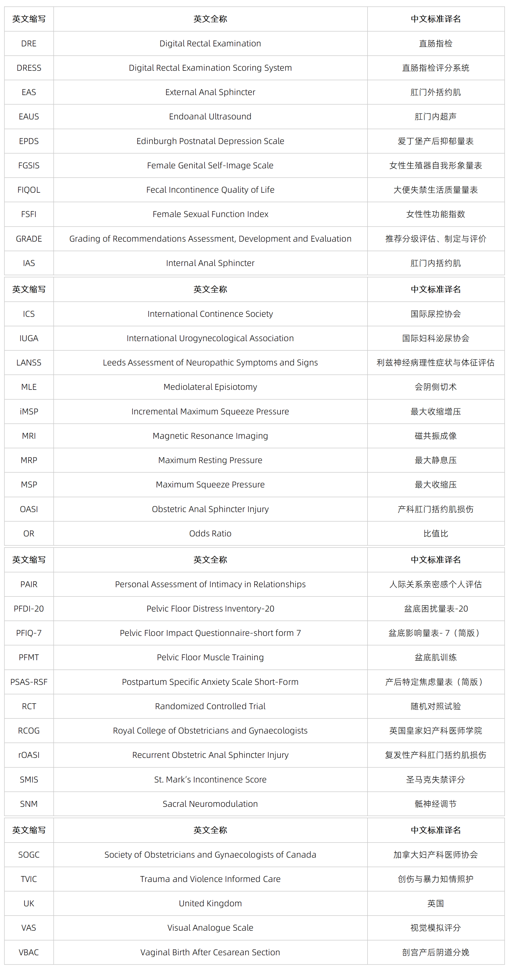
Abbreviation List
Source:
Giroux M, Carter Ramirez A, Dufour S, et al. Guideline No. 465: Obstetrical Anal Sphincter Injuries (OASIs) Part II: Long-Term Management and Counselling Regarding Subsequent Mode of Delivery[J]. Journal of Obstetrics and Gynaecology Canada, 2026, 48(1): 103186. https://doi.org/10.1016/j.jogc.2025.103186
[Translated by Obgyn.net from the "Guideline No. 465: Obstetrical Anal Sphincter Injuries (OASIs) Part II: Long-Term Management and Counselling Regarding Subsequent Mode of Delivery" issued by the Society of Obstetricians and Gynaecologists of Canada (SOGC). For medical professionals only for learning and exchange, not as a basis for specific clinical operations. Actual clinical decisions must be made in conjunction with patient conditions, hospital facilities, and the latest guidelines; multidisciplinary consultation should be conducted when necessary.]
Editor: Lily