





















A Comprehensive Guide to Avoiding Pitfalls in Gynecologic Single-Incision Laparoscopic Surgery: Detailed Strategies for the Prevention and Management of Complications, from Incisional Hernia to Instrument Interference
With the deepening of the minimally invasive concept and the rapid development of technology, laparoendoscopic single-incision surgery (LESS) has gained widespread application in the field of gynecology. This technique is performed through a single incision (transumbilical or transvaginal), maximizing the reduction or concealment of surgical scars, while also offering advantages such as less postoperative pain, faster recovery, and shorter hospital stays. Currently, LESS has been successfully applied to various procedures, including ectopic pregnancy, ovarian cysts, uterine fibroids, total hysterectomy, and even radical surgery for early-stage gynecologic malignancies. It is highly recognized by both patients and surgeons for its excellent cosmetic outcomes and the convenience of specimen extraction.
However, single-incision surgery is by no means simply a "reduction of ports" from multiport laparoscopy. Studies indicate that when the incision is located within or around the umbilicus, the incidence of postoperative incisional hernia in LESS is approximately three times that of traditional multiport laparoscopy. All operating instruments and the camera enter the abdominal cavity through a single channel, breaking the traditional "surgical triangle" of laparoscopy. The limited space affects the surgeon's judgment of the visual field depth, and the mutual interference between instruments, known as the "chopstick effect," further increases the surgical difficulty and learning curve, becoming a core technical challenge that restricts surgical fluency. This article systematically elaborates on the prevention strategies and key management points for complications in gynecologic LESS, combining the latest clinical evidence and operational experience.
Prevention and Management of Incisional Hernia
1. Anatomical Basis and Risks
The umbilicus is a congenital weak point of the abdominal wall. This area lacks muscle layer coverage, has relatively thin subcutaneous fat, and is composed of skin, dense connective tissue (linea alba), and peritoneum, resulting in limited tensile strength. In LESS, to reduce operative difficulty and facilitate specimen extraction, a 2-3 cm fascial incision is typically required, which compromises the integrity of the umbilical ring. Further enlargement and traction of the incision during the operation can increase tension on the fascial edges, leading to ischemic injury and elevating the risk of incisional hernia.
2. High-Risk Factors
According to UpToDate and related clinical guidelines, identifying and managing high-risk factors is key to preventing incisional hernia, including:
• Patient-related factors: Obesity (BMI >30 kg/m²), diabetes, advanced age (>60 years), chronic cough, history of constipation, connective tissue diseases, etc.
• Surgery-related factors: Incision size and closure technique, improper suturing technique, prolonged operation time, sustained intraoperative increase in intra-abdominal pressure (>15 mmHg).
• Postoperative factors: Abdominal distension, vomiting, early strenuous activity, incision infection, etc.
3. Stratified Prevention Strategies
(1) Incision Protection
Small specimens can be extracted through the port channel. For large specimens or those suspicious for malignancy, the concurrent use of a wound protector/retractor is recommended during extraction. It evenly distributes the tension on the incision edges, preventing fascial tearing from excessive traction, and strengthens protection. It serves as the first line of defense against incisional hernia, incision infection, and tumor seeding.
(2) Fascial Closure Technique (Of Paramount Importance)
• Suture Material Selection: Theoretically, the use of slowly absorbable or non-absorbable monofilament sutures (e.g., PDS II or Prolene) is recommended to provide adequate long-term support. In clinical practice, for convenience, 1/2 circle 2-0 absorbable barbed sutures are often used. For obese patients, reinforced sutures can be selected.
• Suturing Technique: Use small skin retractors (12.5 cm) to expose the edges of the anterior rectus sheath and peritoneum on both sides. Perform "figure-of-eight" sutures at both ends and the midpoint of the incision to ensure accurate apposition of the full-thickness fascia, including the peritoneum. The first stitch is placed 0.5-1.0 cm outside the fascial edge on one side, passing through the corresponding position on the opposite side. For longer incisions, continuous suturing can be used; the ratio of suture length to incision length should be ≥4. The suture width and spacing should each be controlled at about 5 mm, known as the "small-bite" technique. The suture width should not exceed 10 mm, otherwise it increases the risk of tissue ischemia and incisional hernia.
• Key Operational Points: Suturing must be performed under direct visualization with clear exposure, confirming that the bowel or omentum is not grasped.
• For Obese Patients: A retractor or small right-angle forceps can be used to push aside the subcutaneous fat to expose the fascial edges before suturing, or a dedicated fascial closure device can be utilized.
(3) Postoperative Management
• Intra-abdominal Pressure Control: Actively manage postoperative cough, constipation, nausea, and vomiting. Instruct patients to avoid lifting heavy objects for 3 months postoperatively.
• Use of Abdominal Binder: For high-risk patients, an elastic abdominal binder can be used postoperatively, primarily to provide external support and alleviate pain.
• Management of Underlying Conditions: Strictly control blood glucose, encourage smoking cessation, and improve nutritional status.
4. Management of Incisional Hernia
Once an incisional hernia occurs, the treatment plan should be determined based on its size, symptoms, and impact on the patient's quality of life. Small, asymptomatic hernias can be observed. Symptomatic or progressively enlarging hernias are recommended for surgical repair.
Summary and Management of Other Complications
1. Bleeding and Hematoma
• Anatomical Variation and Prevention: There are individual differences in the distribution of periumbilical vessels. Using an open technique to establish pneumoperitoneum and performing the fascial incision under direct visualization is recommended.
• Management of Postoperative Hematoma: Small hematomas (diameter <3 cm) can be managed conservatively with observation and local pressure dressing. For progressively enlarging or significantly painful hematomas, ultrasound evaluation is needed. If necessary, sutures should be removed to evacuate the hematoma and achieve hemostasis again.
2. Infection and Umbilical Wound Healing
The umbilicus is a natural site for harboring bacteria, leading to a relatively higher risk of postoperative infection. Preventive measures include: thorough preoperative cleaning of the umbilicus (patients can be instructed to clean in advance with a cotton swab dipped in soap or diluted povidone-iodine); strict aseptic technique during surgery, using a wound protector; keeping the incision dry and clean postoperatively. Once infection occurs, antibiotic treatment should be administered based on severity. If an abscess forms, incision and drainage are required.
3. Specimen Extraction Complications
Specimen extraction is one of the challenges in LESS, especially for larger or nature-uncertain masses. To avoid spillage of contents from cyst rupture (particularly endometriotic cysts and potential tumor seeding), the principle of "in-bag manipulation" must be adhered to. All excised tissues should be placed inside a retrieval bag before further handling. For larger uterine fibroids or the uterus, morcellation can be performed inside the bag, akin to "apple-coring," ensuring the cutting instrument tip is always kept in view within the bag. If rupture occurs, the incision should be immediately irrigated, the retrieval bag replaced, and the incision extended if necessary for complete specimen removal.
Prevention and Overcoming of Instrument Interference ("Chopstick Effect")
The "chopstick effect" is the most core technical challenge in LESS, referring to the mutual interference between the camera and operating instruments within the same channel, leading to the loss of the surgical triangle, restricted range of motion, and limited field of view.
1. Hardware Solutions
• Instrument Selection:
◦ Flexible/Pre-bent Instruments: Pre-bent shaft design, allowing the handle to deviate from the operating axis, avoiding collision outside the body.
◦ Articulating Instruments: Tip allows multi-angle articulation, facilitating fine manipulation in confined spaces.
◦ Extended-length Instruments: Increase the length of the operating shaft, moving the handles out of the mutual interference zone. (Conventional instrument length 330mm, extended length 420mm)
• Lens Selection:
◦ 10mm High-Definition Lens: The light source is located on the main body of the lens, reducing the space occupied by the light cable.
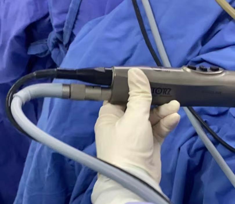
◦ 3D High-Definition Lens: Provides stereoscopic vision, compensating for the loss of depth perception in single-incision operations, reducing operative difficulty.
◦ 5mm 30° High-Definition Extra-long Lens: The viewing angle can be adjusted by rotating the lens, avoiding instrument obstruction of the view; to some extent reduces interference between the lens and surgical instruments, increasing the stability of the operation. (Specifications: 5mm*500mm)
• Lens Optimization: Using a 3D laparoscope can provide better depth perception, aiding in performing delicate operations within a crowded space.
2. Operative Skills and Team Coordination
• Crossing Technique: This is the core maneuver to overcome the "chopstick effect." The left-hand instrument operates the tissue on the right side, and the right-hand instrument operates the tissue on the left side, creating an instrument crossover inside the abdominal cavity, thereby providing more working space externally.
• Camera Holder Coordination: The camera holder must possess the awareness of a "third eye," actively avoiding the surgeon's operating instruments, and proactively adjusting the view based on the surgical progress, always keeping the key anatomical structures in the center of the field of view.
• Positioning Adjustment:
◦ The Trendelenburg position utilizes gravity to displace the intestines out of the pelvis, reducing the need for instrument swinging.
◦ Lateral tilt: When operating on the adnexa of one side, tilt the operating table 15-20° towards the opposite side, suspending the target organ at the highest point for better exposure.
◦ Dynamic adjustment: Make real-time fine adjustments to the patient's position based on the surgical steps, rather than keeping it fixed.
Camera Holder Techniques:
The camera holder needs to establish默契配合 (tacit cooperation) with the surgeon:
• View Anticipation: Adjust the lens direction in advance, keeping the center of the view always directed at the surgeon's target area.
• Avoidance Maneuver: When the surgeon's instrument moves, the camera holder slightly retracts the lens, following and zooming in only after the instrument is in position.
• Stability Priority: The operating space in single-incision surgery is limited; lens shaking can cause significant visual dizziness. The principles of "steady, accurate, and slow" are emphasized.
Conclusion
Gynecologic single-incision laparoscopic surgery is a paradigm combining minimally invasive concepts with aesthetic pursuit. However, its safe implementation relies on a profound understanding and systematic prevention of complications. Successful LESS surgery depends on the philosophy of "prevention over treatment." By strictly selecting appropriate indications, identifying and managing high-risk factors, adopting refined fascial suturing techniques (such as the "small-bite" continuous suture), and mastering the skills to overcome instrument interference, the risk of complications can be minimized.
References:
[1] Gynecologic Single-Incision Laparoscopic Surgery (including vNOTES) Expert Consensus Group, Chinese Society of Obstetrics and Gynecology, Chinese Medical Association. Expert consensus on gynecologic single-incision laparoscopic surgery (including vNOTES) (2025 Edition). Chinese Journal of Obstetrics and Gynecology, 2025, 60(6): 417-421.
[2] Antoniou SA, Morales-Conde S, Antoniou GA, et al. Single-incision laparoscopic surgery through the umbilicus is associated with a higher incidence of trocar-site hernia than conventional laparoscopy: a meta-analysis of randomized controlled trials. Hernia 2016; 20:1.
[3] Yang Y, Pan YQ, Lu Q, Bao W, Wang M, Liu W, Wu SF. Ergonomic learning curves on gynecological laparoendoscopic single-site (LESS) surgery. BMC Surg. 2023 Oct 27;23(1):327. doi: 10.1186/s12893-023-02241-x. PMID: 37891581; PMCID: PMC10612358.
[4] Itatsu K, Yokoyama Y, Sugawara G, et al. Incidence of and risk factors for incisional hernia after abdominal surgery. Br J Surg 2014; 101:1439.
[5] Clinical features, diagnosis, and prevention of incisional hernia. UpToDate. Literature review current through: 2025-05-24.
[6] Principles of abdominal wall closure. UpToDate. Topic last updated: 2025-03-19.
[7] Complications of minimally invasive surgery. UpToDate. Topic last updated: 2025-08-21.
[8] Access techniques for minimally invasive surgery. UpToDate. Topic last updated: 2025-08-12.
Author Introduction

Image source: Provided by the author of this article.
Responsible editor: Lily