





















"Incurable Cancer" Adenomyosis: What Are the Preferred Uterus-Preserving Treatment Options Beyond Hysterectomy?
As a gynecologist, I am often asked a heart-wrenching question by my patients: "Doctor, is removing my uterus the only way to cure this disease?"
The women who ask this are often those who have suffered from adenomyosis for years. Among them, some experience menstrual pain so severe it prevents them from working, others suffer from such heavy bleeding that they develop significant anemia, and many have sought advice from multiple hospitals only to hear the same suggestion: "Just have a hysterectomy; it's a permanent solution."
Adenomyosis, a benign gynecological condition often called an "incurable cancer" – does it really have to end with the removal of the uterus? Today, I'd like to discuss the preferred uterus-preserving treatment options, based on the latest clinical guidelines and case studies.
Precise Classification of Adenomyosis
The first step in uterus-preserving treatment is precise classification. Treatment strategies differ drastically for different types of adenomyosis. Based on MRI imaging features, the Kishi classification is the most commonly used in clinical practice:
• Type I (Internal/Endometrial): Lesions originate from the endometrial-myometrial junctional zone (JZ). The JZ is thickened, and the outer myometrium is not affected.
• Type II (External/Subserosal): Lesions affect only the outer layer of the uterus. The JZ remains intact, with normal myometrial tissue visible between the JZ and the lesion.
• Type III (Intramural): Lesions are isolated, surrounded by intact myometrium, and show no continuity with either the JZ or the serosal layer.
• Type IV (Diffuse/Unclassified): Lesions are widespread and diffuse, making it impossible to clearly identify their point of origin.
Why is Classification So Important?
Different types of adenomyosis have significantly different impacts on fertility and pregnancy outcomes. Patients with the diffuse type primarily experience adverse obstetric outcomes, with higher rates of second-trimester miscarriage, premature rupture of membranes, gestational hypertension, and intrauterine infection compared to those with focal adenomyosis. Furthermore, the risk of miscarriage and cervical insufficiency increases as lesion size grows. Focal adenomyosis is more closely associated with primary infertility, with the risk of infertility increasing 1.9-fold when the outer myometrium is involved. Larger lesions correlate with lower live birth rates. The internal type (especially with JZ involvement) carries a threefold increased risk of miscarriage after embryo transfer, with significantly lower clinical pregnancy rates. Severe cases have a miscarriage rate double that of the normal population. While the external type has a relatively lower miscarriage risk, it is associated with a higher risk of abnormal placental location during pregnancy. Additionally, increased uterine volume, asymmetric myometrial thickening, and abnormal uterine cavity morphology all indicate poorer pregnancy outcomes. Therefore, it is recommended that patients undergo pelvic MRI (which is more accurate than ultrasound) to confirm the classification before proceeding with uterus-preserving treatment.
Preferred Uterus-Preserving Option 1: Medication – Not Merely Symptom Management
Many patients, upon hearing "medication," think it's just buying time. In reality, modern drug therapy is far from simple pain relief or bleeding control; it represents targeted intervention based on the disease mechanism.
1. GnRH-a: The "Golden Standard" for Pre-treatment
Gonadotropin-releasing hormone agonists (GnRH-a) work by suppressing ovarian function, creating a "pseudo-menopausal" state that causes ectopic endometrial tissue to atrophy.
• Key Applications: Used in long or ultra-long protocols for 3-6 months of pre-treatment. This can significantly reduce uterine volume, creating favorable conditions for subsequent surgery or assisted reproduction. It's crucial to use "add-back therapy" (low-dose estrogen + progestin) alongside GnRH-a to maintain efficacy while alleviating hypoestrogenic side effects like hot flashes and bone loss. GnRH-a is also used as adjuvant therapy after conservative surgery. Evidence suggests that using GnRH-a after conservative surgery can significantly increase pregnancy rates, with natural conception rates reaching 40.7%, compared to only 15.0% in patients who do not receive it.
2. Dienogest: A New Choice for Long-Term Management
This novel selective progesterone receptor modulator is taken orally at a recommended dose of 2 mg/day, continuously. Its characteristic is that it suppresses endometrial proliferation without completely inhibiting ovulation. For patients with no immediate fertility plans who wish for long-term uterus preservation, this is a gentler option compared to GnRH-a. Note: Be aware of the potential side effect of irregular vaginal bleeding, which is common in the first 3-6 months but is tolerable for most.
3. LNG-IUS (Mirena): An Underestimated Tool
The levonorgestrel-releasing intrauterine system (LNG-IUS) is primarily used to treat dysmenorrhea, chronic pelvic pain, and menorrhagia. It works by releasing a high local concentration of progestin directly onto the endometrial-myometrial junctional zone, achieving a triple effect: reducing menstrual flow, alleviating pain, and inhibiting lesion progression. Indications: Suitable for women who have completed childbearing or have no immediate fertility plans; for use as postoperative maintenance therapy to reduce recurrence risk; and for endometrial preparation prior to assisted reproduction (3 months of use can significantly improve embryo implantation rates). For patients with significantly enlarged uteri or severe pain, a sequential approach can be used: first, apply GnRH-a for 3-6 months to reduce uterine size, then insert the LNG-IUS. This lowers the risk of LNG-IUS expulsion.
Preferred Uterus-Preserving Option 2: Conservative Surgery – More Complex Than Just "Removing a Tumor"
When medication is ineffective or there are clear focal lesions, lesion excision surgery (while preserving the uterus) is an important option. However, it must be emphasized: surgery for adenomyosis is far more challenging than myomectomy (fibroid removal). The Chinese Expert Consensus on the Diagnosis and Management of Adenomyosis clearly states: For adenomyosis with infertility, conservative surgery is not recommended as first-line treatment. It is suggested as an alternative for patients with severe symptoms or repeated ART (assisted reproductive technology) failure, and it yields better results in cases of focal adenomyosis.
1. Focal Lesion Excision (Adenomyomectomy)
Applicable to patients with Kishi Type II or III. Surgical Key Points: Lesion Delineation – The boundary between adenomyosis lesions and normal myometrium is often indistinct, making preoperative MRI precise localization crucial. Suturing Technique – Multi-layer closure of the myometrium is essential to restore anatomical structure and reduce the risk of uterine rupture during pregnancy. Intraoperative Adjuncts – Combined hysteroscopic and laparoscopic approaches ensure a normal uterine cavity shape. Efficacy Data: A systematic review cited in the Chinese Expert Consensus shows a postoperative pregnancy rate of 52.7% after focal adenomyosis lesion excision, significantly higher than the 34.1% rate for diffuse adenomyosis.
2. "Volume Reduction" Surgery for Diffuse Adenomyosis
For diffuse adenomyosis, complete lesion removal is almost impossible (equivalent to removing the entire uterine wall). In such cases, lesion volume reduction surgery combined with uterine reconstruction, followed by long-term medication management, can be employed. Important Reminder: Conservative surgery carries a high risk of serious complications like uterine rupture during pregnancy and placenta previa. Postoperative contraception for 6-12 months is mandatory, and close monitoring of the uterine scar during pregnancy is essential.
Preferred Uterus-Preserving Option 3: Minimally Invasive Ablation – The "Invisible Scalpel"
This category includes Uterine Artery Embolization (UAE), High-Intensity Focused Ultrasound (HIFU), and radiofrequency or microwave ablation. In clinical application, as interventional or physical therapies can only reduce lesion size and improve symptoms but cannot excise the lesion or provide tissue for pathological examination, thorough patient evaluation and strict adherence to indications are required before treatment. This is especially important to exclude patients with malignancy or suspected malignancy.
• ① UAE Indications: Acute uterine bleeding due to adenomyosis; medication failure; patient refusal of or inability to tolerate surgery. Strict adherence to indications is crucial, especially to exclude malignancy.
• ② HIFU Ablation Indications: Symptomatic adenomyosis; myometrial thickness at the lesion site >3 cm; premenopausal women; successful targeting with on-board imaging and adequate acoustic pathway. Advantages: Non-invasive, preserves uterine myometrial structure, allows pregnancy planning 3-6 months post-procedure. Patients with the internal type (Type I) show the best efficacy, with pregnancy rates reaching 59.6%. Treatment can be repeated if recurrence occurs, making it an ideal choice balancing symptom relief and fertility preservation.
• ③ Radiofrequency or Microwave Ablation: While these methods have established efficacy in treating uterine fibroids, their clinical application in adenomyosis is less common and requires further research.
Adenomyosis is indeed challenging, but it is by no means a condition where "only hysterectomy is the answer." As doctors, our duty is not only to treat the disease but also to help patients find the best balance between efficacy and quality of life. Preserving the uterus preserves not just an organ, but a woman's dignity and hope.
References:
[1] Wang Shixuan, Cui Pengfei, Zhang Jinjin. Expert Consensus on Three-Tier Management of Adenomyosis. Journal of Practical Obstetrics and Gynecology, 2024, 40(02): 106-111.
[2] Chinese Association of Obstetricians and Gynecologists, Endometriosis Collaborative Group of Chinese Society of Obstetrics and Gynecology. Guidelines for Diagnosis and Treatment of Endometriosis (Third Edition). Chinese Journal of Obstetrics and Gynecology, 2021, 56(12): 812-824.
[3] Huang Yan, Peng Chao, Zhou Yingfang. Diagnosis, Treatment, and Management of Pain Related to Adenomyosis. Chinese Journal of Practical Gynecology and Obstetrics, 2025, 41(08): 784-790.
[4] Ouyang Zhenbo, Zhong Biting, Liu Minyin, et al. Interpretation of the Asian Endometriosis and Adenomyosis Society Guidelines for Adenomyosis Management. Obstetrics, Gynecology and Genetics (Electronic Edition), 2024, 14(01): 1-5.
[5] Endometriosis Committee, Chinese Association of Obstetricians and Gynecologists. Chinese Expert Consensus on Diagnosis and Management of Adenomyosis. Chinese Journal of Obstetrics and Gynecology, 2020, 55(06): 376-383.
[6] Dason E Shirin, et al. Guideline No. 437: Diagnosis and Management of Adenomyosis. Journal of Obstetrics and Gynaecology Canada, 2023.
[7] Pang Li Li, et al. Efficacy of High-Intensity Focused Ultrasound Combined With GnRH-a for Adenomyosis: A Systematic Review and Meta-Analysis. Frontiers in Public Health, 2021.
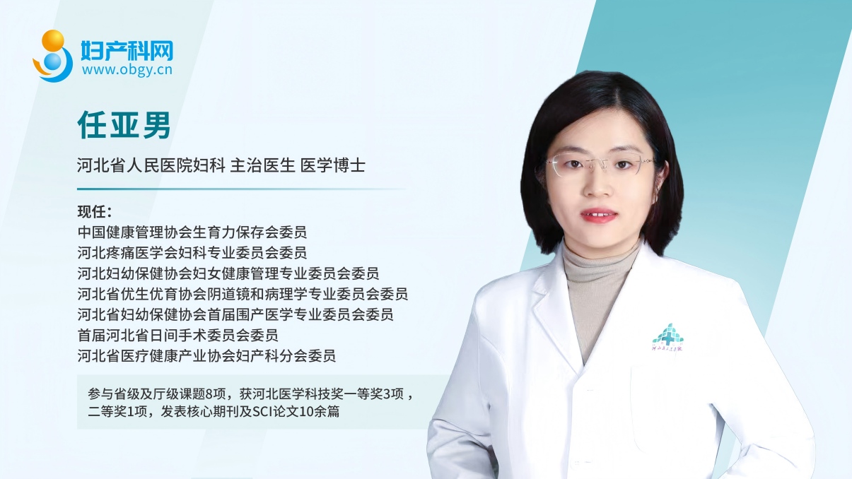
About the Author
Editor: Huo Pan