





















Ectopic pregnancy is a life-threatening reproductive health risk for women of childbearing age. Its global incidence continues to rise, with special subtypes like cesarean scar pregnancy becoming increasingly prominent, easily leading to severe complications such as fatal hemorrhage, especially in low- and middle-income countries. Although advancements in technologies like laparoscopic surgery and high-resolution ultrasonography have driven diagnostic and therapeutic progress, challenges remain, including the risk of misdiagnosis for rare subtypes, controversies in treatment selection (e.g., expectant management vs. surgery), and new challenges arising from assisted reproductive technology. This article synthesizes core evidence-based content from a significant Grand Theme Review published in 2026 in Human Reproduction Update.
(Image source: Hum Reprod Update)
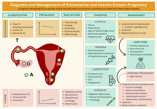
The review systematically compiled the latest global evidence up to July 2025, covering the standardized terminology and classification, epidemiology and risk factors, precise diagnostic pathways based on ultrasound and biochemical markers, evidence-based recommendations for individualized treatment options (including expectant management, medication, and surgery), and key points for long-term care and psychological support for ectopic pregnancy.
I. Definitions and Terminology Standardization
Ectopic pregnancy refers to a pregnancy implanted completely or partially outside the endometrial cavity, which may lead to hemorrhage-related complications due to growth in an abnormal anatomical location. Terminology consensuses were established by the European Society of Human Reproduction and Embryology (ESHRE) in 2020 and the Society of Radiologists in Ultrasound (SRU) in 2025, with core specifications as follows:
1. Normal Pregnancy
ESHRE uses "normally implanted pregnancy." SRU may use "normally located pregnancy" or "normally located intrauterine pregnancy," both referring to a pregnancy within the endometrial-myometrial junction zone. The term "cornual pregnancy" is obsolete.
2. Ectopic Pregnancy Classification
◦ Extrauterine Ectopic Pregnancies: Include tubal, ovarian, abdominal, and interstitial pregnancies.
◦ Intrauterine Ectopic Pregnancies: Include cesarean scar, cervical, intramural, and pregnancies associated with uterine malformations.
◦ Special Types: Partial (pregnancy straddling anatomical boundaries), complete (pregnancy entirely confined to an abnormal site), and heterotopic (concurrent normal and ectopic pregnancies).
3. Pregnancy Status Description
ESHRE uses "viable" (fetal cardiac activity visible) and "failing" (spontaneous resolution suggested by ultrasound or hormones). SRU advises against using the term "viable."
II. Incidence and Mortality
1. Incidence
The global age-standardized incidence in 2019 was 170 per 100,000 women, with the 25-29 age group at highest risk. The peak shifts to ages 30-34 in high sociodemographic index regions. Ectopic pregnancy rates associated with Assisted Reproductive Technology (ART) vary geographically and over time. A UK cohort study (2000-2012) reported an incidence of 1.4%. US data (2001-2011) showed a declining trend, from 2.0% to 1.6%. Single embryo transfer carries a relatively lower risk, while transferring 4 or more embryos increases the risk.
2. Proportion of Specific Types
Tubal pregnancies account for 95%–97%. Other extrauterine sites (ovarian, abdominal, etc.) account for about 1%–2%. The incidence of intrauterine ectopic pregnancies like cesarean scar pregnancy is rising.
3. Mortality
The global age-standardized mortality in 2019 was 0.16 per 100,000 women, significantly higher in low- and middle-income countries. The maternal mortality rate in the UK was 0.4 per 100,000 maternities in 2018-2020, rising to 0.8 per 100,000 in 2021-2022. Women from ethnic minorities face higher complication risks.
III. Risk Factors, Etiology, and Pathogenesis
(A) Risk Factors
1. Tubal Ectopic Pregnancy:
Core risk factors include tubal damage (infection, endometriosis, pelvic surgery history), smoking, advanced maternal age, infertility history, and prior ectopic pregnancy (recurrence risk 5%–19%). Among women with an intrauterine contraceptive device (IUCD) who become pregnant, ectopic risk is elevated; low-dose levonorgestrel IUCDs carry the highest risk (2.76/1000 woman-years). The adjusted odds ratio for risk associated with levonorgestrel-based emergency contraception is 4.75-10.5.
2. Other Extrauterine Ectopic Pregnancies:
Ovarian and abdominal pregnancies are associated with tubal pathology; IUCD use is strongly linked to ovarian ectopic pregnancy (OR=9.6).
3. Intrauterine Ectopic Pregnancies:
◦ Cesarean Scar Pregnancy: Requires the presence of a myometrial defect (niche). Multiple cesarean sections and a retroverted uterus are high-risk factors.
◦ Intramural Pregnancy: Associated with endometrial-myometrial junction damage (e.g., myomectomy, uterine perforation).
◦ Cervical Pregnancy: Risk factors include multiple abortions, curettage history, and smoking (OR=2.82).
4. ART-Related Risks:
ART can increase the risk of all ectopic types, associated with the number of embryos transferred, tubal factor infertility, thin endometrium, day-3 cleavage-stage embryo transfer, and diminished ovarian reserve. Frozen-thawed embryo transfer and natural cycle IVF carry relatively lower risks.
(B) Etiology and Pathogenesis
1. Tubal Pregnancy:
Elevated progesterone and inflammatory factors like interleukin-6 can reduce tubal ciliary beat frequency. Smoking increases risk by affecting smooth muscle contraction. Infections like chlamydia can upregulate integrin β1, promoting embryo adhesion to tubal epithelium.
2. Intrauterine Ectopic Pregnancy:
Inflammatory responses, reduced myometrial volume, and a hypoxic environment at the cesarean scar can stimulate trophoblast invasion. Cervical and intramural pregnancies often result from surgical damage causing anatomical disruption, allowing implantation outside the endometrial cavity.
IV. Clinical Assessment and Diagnosis
(A) Clinical Assessment
1. Common Symptoms:
Bleeding and abdominal pain are primary presentations. Cesarean scar and cervical pregnancies may present as painless vaginal bleeding. Ovarian pregnancy is more likely to present as abdominal pain alone, even hemorrhagic shock (~7%). Bleeding lasting >3 days increases the daily risk by 20% (95% CI 14%–27%).
2. Critical Signs:
Hemodynamically unstable presentation (e.g., postural hypotension) in a woman of childbearing age suggests potential massive intra-abdominal hemorrhage. In LMICs, culdocentesis can assist in confirming intra-abdominal bleeding.
(B) Imaging Diagnosis
1. Transvaginal Ultrasound (TVS):
The first-line investigation, capable of determining pregnancy location, morphological features, and intra-abdominal hemorrhage. Detection rates in expert centers are 74%–83%. Operator-dependent; uncertain cases should be referred to specialized centers.
2. Ultrasonographic Features by Type:
◦ Tubal Pregnancy: Presents as a heterogeneous mass or a structure containing a gestational sac, yolk sac/embryo. The "interstitial line sign" may be seen in interstitial pregnancies.
◦ Cesarean Scar Pregnancy: Gestational sac located within the scar defect, with thinning of the myometrium between the sac and bladder wall, rich vascularity; placental lacunae visible in 44%.
◦ Abdominal Pregnancy: The gestational sac can be anywhere in the peritoneal cavity. In later stages, the fetus may be seen adjacent to the abdominal wall, with the placenta outside the uterine cavity.
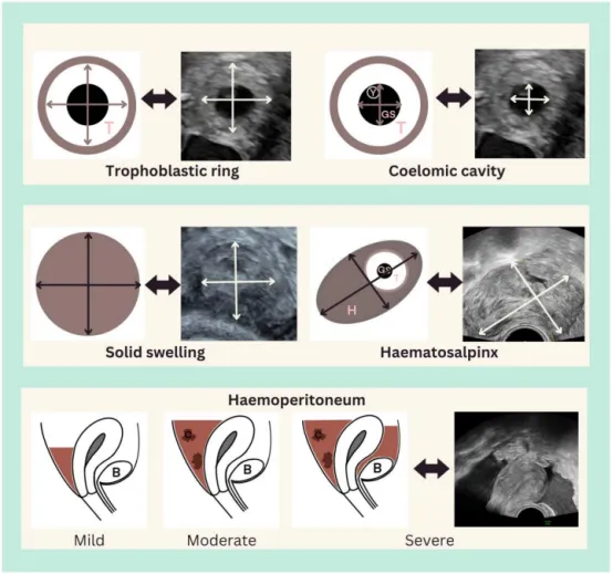
Note: Ultrasound measurements for tubal ectopic pregnancy and hematosalpinx, and assessment of hemoperitoneum. All measurements are presented in 2D; a third orthogonal dimension measurement is also needed. Hyperechoic trophoblast (T) is visible within the tubal lumen, which may contain a gestational sac (GS). Surrounding hematosalpinx (H) may be present. If structures like a yolk sac (Y) or embryo are visible within the sac, they should be measured similarly to a normal pregnancy. Clotted blood in the pelvis suggests moderate hemoperitoneum, while hemorrhage within the vesicouterine pouch (B) suggests severe hemoperitoneum.
3. Hemorrhage Assessment:
Mild: anechoic fluid in the pouch of Douglas; Moderate: presence of blood clots; Severe: clots extending to the vesicouterine pouch. Presence of fluid in the hepatorenal recess suggests blood loss ≥670 ml.
(C) Biochemical Assessment
1. Human Chorionic Gonadotropin (hCG):
No single "safe" threshold excludes ectopic pregnancy. A single measurement has limited value; 10%–20% of ectopic pregnancies show hCG patterns similar to normal pregnancy, thus it cannot be used alone for diagnosis.
2. Progesterone:
Can identify low-risk failing pregnancies. When progesterone ≤10 nmol/L, the risk of intervention is only ~2%, but it cannot exclude a viable normal pregnancy. In IVF patients, progesterone levels are unreliable due to exogenous supplementation.
3. Pregnancy of Unknown Location (PUL):
Refers to cases where ultrasound does not confirm pregnancy location, accounting for 7%–47% of early pregnancy assessments.
(D) Surgical and Histological Assessment
1. Surgical Assessment:
Laparoscopy is rarely the first-choice diagnostic method, reserved for hemodynamically unstable cases or those with high suspicion, with a false-positive rate of 0.6%. Severe pelvic adhesions may lead to false negatives.
2. Histological Assessment:
Surgical specimens require histological examination for definitive diagnosis; about 5% of surgically confirmed ectopic pregnancies yield negative histology. Extravillous trophoblast proliferation is more pronounced in tubal pregnancies, posing a risk of overdiagnosis as molar pregnancy.
V. Treatment Options
Core principles: Avoid overtreatment, reduce iatrogenic harm while ensuring maternal safety. Treatment must be individualized based on pregnancy type, status (viable/failing), location, and patient's fertility desires.
(A) Tubal Ectopic Pregnancy
1. Expectant Management:
Suitable for asymptomatic patients with hCG <1000 IU/L, pregnancy mass <35mm, and no viable embryo. Success rate 31%–40%. May be cautiously considered for hCG 1000-1500 IU/L. Average follow-up 15 days; 95% of masses resolve on ultrasound within 78 days.
2. Medical Treatment:
Primarily methotrexate (a folic acid antagonist). Suitable for stable patients without a viable embryo and hCG <5000 IU/L. Dose: 50mg/m². An hCG decline <15% between days 4-7 requires an additional dose. For tubal ectopic pregnancy (TEP) with hCG 1000-5000 IU/L, median time to biochemical resolution is 28 days, 14% require an additional dose, and 29% ultimately require surgery. Note: Methotrexate has limited efficacy for intrauterine ectopic pregnancies like cesarean scar pregnancy and is not recommended as first-line treatment.
3. Surgical Treatment:
Laparoscopic surgery is preferred. Salpingectomy is the first-line option for hemodynamically unstable patients. Based on the ESEP RCT, the ongoing pregnancy rate at 3 years after salpingostomy (61%) showed no statistically significant difference compared to salpingectomy (56%). However, salpingostomy carries a significantly higher risk of persistent trophoblast (7% vs. <1%) and a non-significantly higher recurrence risk (8% vs. 5%). Additionally, about 20% of patients undergoing salpingostomy require a second surgery for salpingectomy due to persistent bleeding.
(B) Other Extrauterine Ectopic Pregnancies
1. Ovarian Pregnancy:
Primarily treated with laparoscopic surgery, involving pregnancy tissue resection or ovarian wedge resection. Median intraoperative blood loss is 700ml. Postoperative hCG monitoring is needed to rule out persistent trophoblast.
2. Abdominal Pregnancy:
Often requires surgery. Preoperative localization of the implantation site is crucial, with preparation for massive transfusion. If the placenta is densely adherent, it may be left in situ, with reassessment for potential secondary surgery at 3 months.
(C) Intrauterine Ectopic Pregnancies
1. Cesarean Scar Pregnancy:
◦ Expectant Management: Carries a very high risk for cesarean scar pregnancies with fetal cardiac activity. A systematic review showed ~13% of such pregnancies result in severe vaginal hemorrhage, with up to 15% requiring hysterectomy in the first or second trimester. As these are essentially precursors to the placenta accreta spectrum, with a high risk of progressing to mid-late pregnancy, routine expectant management is not recommended.
◦ Medical Treatment: Systemic methotrexate success rate is only 59%, local injection ~74%, but resolution time is long (median 88 days).
◦ Surgical Treatment: Ultrasound-guided transcervical suction evacuation is the main method. Complications are lower in patients treated before 9 weeks, with crown-rump length <23mm, and low vascularity.
2. Cervical Pregnancy:
Treatment principles are similar to cesarean scar pregnancy. Based on case series literature, combined surgical evacuation and uterine artery embolization yields a comprehensive success rate of ~87%, with ~9% of patients requiring hysterectomy.
3. Intramural Pregnancy:
Partial types may be treated with transcervical suction evacuation. Complete types require laparoscopic/open surgery to remove the pregnancy tissue and repair the myometrium. Expectant management can be considered if hCG is declining; median time to biochemical resolution is 71 days, and ultrasound resolution is 214 days.
VI. Long-term Care
(A) Management of Continuing Pregnancy
If a viable ectopic pregnancy is continued, monitoring by a specialist obstetric team is essential, informing of risks like uterine rupture, massive hemorrhage, and extreme prematurity. Planned cesarean section at 34-35⁺⁶ weeks is advised. 75% may have placenta accreta, and 58% may require hysterectomy at the time of cesarean section.
(B) Subsequent Pregnancy-Related
1. Fertility Prognosis:
70% of women with a prior ectopic achieve a normally implanted pregnancy; 57% achieve a live birth. After unilateral salpingectomy, 32% of pregnancies originate from ovulation on the same side.
2. Recurrence Risk:
Prior ectopic pregnancy increases recurrence risk sixfold. After salpingostomy, recurrence risk is 8% (vs. 5% after salpingectomy). Cesarean scar pregnancy recurrence rate is ~18%.
3. Contraception Advice:
All contraceptive methods reduce the risk of ectopic pregnancy and can be recommended based on patient circumstances.
(C) Psychological and Other Outcomes
1. Psychological Impact:
21% of patients experience post-traumatic stress disorder 9 months post-treatment, 23% have moderate-to-severe anxiety, and 11% have moderate-to-severe depression. Diagnostic uncertainty and pregnancy termination decisions can exacerbate psychological trauma.
2. Cesarean Scar Defect (Niche) Management:
Elective repair can improve abnormal bleeding and infertility, but robust long-term randomized trial evidence is lacking. Repair solely for preventing recurrence is not yet recommended.
VII. Future Research Directions
1. Develop rapid diagnostic models suitable for low-resource settings; optimize assessment pathways for pregnancy of unknown location.
2. Conduct large-sample, long-term follow-up trials to clarify the effectiveness of novel treatments and their impact on fertility and recurrence.
3. Develop a core outcome set to standardize research reporting on ectopic pregnancy.
4. Explore effective psychological interventions to improve psychological outcomes for patients and families.
5. Investigate disparities in ectopic pregnancy incidence and outcomes among ethnic minorities to reduce health inequities.
VIII. Conclusion
Ectopic pregnancy is a significant threat to the reproductive health of women of childbearing age, posing risks of pregnancy loss, life-threatening complications, and impact on future fertility. Timely, precise ultrasound assessment is the cornerstone of safe management. Stable patients should have a confirmed diagnosis before intervention. In clinical decision-making, low-risk patients can be prioritized for expectant management; surgery should be minimally invasive where appropriate. Methotrexate is no longer a first-line treatment for all cases. Long-term care, including fertility counseling and psychological support, is crucial. The future requires standardized management pathways, precise risk-stratified treatment, and multidisciplinary collaboration to further improve patient outcomes.
Content source: Jessica Farren, Bassel H Al Wattar, Davor Jurkovic, The diagnosis and management of extrauterine and uterine ectopic pregnancy, Human Reproduction Update, Volume 32, Issue 1, January-February 2026, Pages 2–32, https://doi.org/10.1093/humupd/dmaf024
[This article is intended for medical professional learning and exchange only and does not constitute specific clinical operation guidance. Actual clinical decisions must be made based on the patient's condition, hospital resources, and the latest guidelines, with multidisciplinary consultation as needed.]
Editor: Lily