





















Global Trends in Cesarean Delivery and the Evolution of Surgical Expertise
According to a January 2026 expert review in the American Journal of Obstetrics and Gynecology, cesarean delivery (CD) is now the most common major surgical procedure globally.
World Health Organization (WHO) data shows the global cesarean delivery rate from 2010-2018 was 21.8%, with vast geographical disparities (5.0%-42.8%).
Experts predict that by 2030, the global average cesarean delivery rate will rise to 28.7%, with East Asia potentially seeing increases exceeding 60%.
In the United States, while the repeat cesarean rate slightly decreased from 32.9% to 31.7% between 2009-2019, the overall cesarean rate has continued to rise since its modern low of 20% in 1996.
Although WHO no longer recommends specific cesarean rate targets, clear evidence indicates that cesarean delivery is associated with an increased risk of severe maternal morbidity compared to vaginal birth. This has driven efforts in various regions to reduce non-medically indicated cesarean deliveries to lower maternal risks. While surgical outcomes have improved through measures like blood transfusion, effective analgesia, and antibiotic prophylaxis, cesarean delivery remains intrinsically a major abdominal surgery associated with higher maternal risks.
☞ A key paradox lies in this:
The complexity of cesarean delivery is increasing, while surgeons' experience in managing complex laparotomies is decreasing. Over the past two decades, the rise of minimally invasive gynecologic surgery has led to a decline in the volume of laparotomies in clinical practice and training programs. Although up to 80% of cesarean deliveries can be safely completed using evidence-based techniques, managing more complex cases (such as those with adhesions after multiple cesareans, large fibroids, uterine rupture, etc.) requires more refined, advanced surgical skills.
This review aims, through descriptions and imagery, to clarify surgical approaches for specific complex cesarean delivery scenarios for frontline physicians.
The "Ten Surgical Truths" for Complex Obstetric Surgery and Traits of Surgical Excellence
The article opens by presenting the "Ten Surgical Truths" and "Traits of Surgical Excellence" that guide complex obstetric surgery, transcending mere technique to emphasize the core importance of decision-making, preparation, and teamwork. (Table 1)
➱ The Ten Truths for Complex Obstetric Surgery include:
1. First, do no harm. Do only what is necessary, but be prepared for anything that is. Practice regularly; have a clear plan for help.
2. Technique trumps technology. Technological advances are tools; sound fundamentals are key.
3. Know your equipment. Particularly for advanced technology, you cannot rely solely on technicians for troubleshooting.
4. Plan thoroughly in advance. Review previous operative notes, prenatal imaging (not just reports), and discuss with the team beforehand.
5. Anatomy is foundational. You must be able to differentiate normal from abnormal anatomy.
6. Go from the easy to the hard, but do not avoid the hard. Like choosing a route home, avoid traffic, but if it's the only way, tell someone and get help.
7. Prioritize clearly. Adjust based on clinical and environmental circumstances. Do not spend hours perfectly dissecting the bladder during fetal distress, nor wait to clamp the cord while the uterine artery extension is bleeding.
8. Work as a team; communicate harmoniously. Communication is critical.
9. Time is blood; time is life. Delays in recognition and response remain a cause of preventable severe morbidity and mortality.
10. Balance confidence with humility. Even the most experienced surgeons encounter unique challenges. Ask for help before becoming entrenched; learn from the challenge.
➱ The Traits of Surgical Excellence are summarized as the 4 A's:
1. Ability: Technical skills (mastery of procedures) and tactical wisdom (judgment on when to act, when to use restraint). Ideally, ability is not static but evolves with skill acquisition and practice, and sometimes declines with loss of function (e.g., after illness or accident). Accurate assessment of one's own and the team's ability is equally crucial.
2. Adaptability: Includes embracing lifelong learning, fostering personal and professional development, introducing surgical approaches based on new evidence, and testing innovative surgical solutions.
3. Availability: Surgery is a hands-on skill requiring both technique and physical presence. Similarly, being present from the initial preoperative visit through postoperative care allows surgeons to anticipate, recognize, manage, and learn from complications early.
4. Affability and humility: Our work is challenging. Managing stress by laughing at trivialities and finding joy in simple pleasures helps maintain a sense of purpose during difficult times. Maintaining a growth mindset and recognizing that we will not see 'everything' before retirement drives continuous learning from every case.
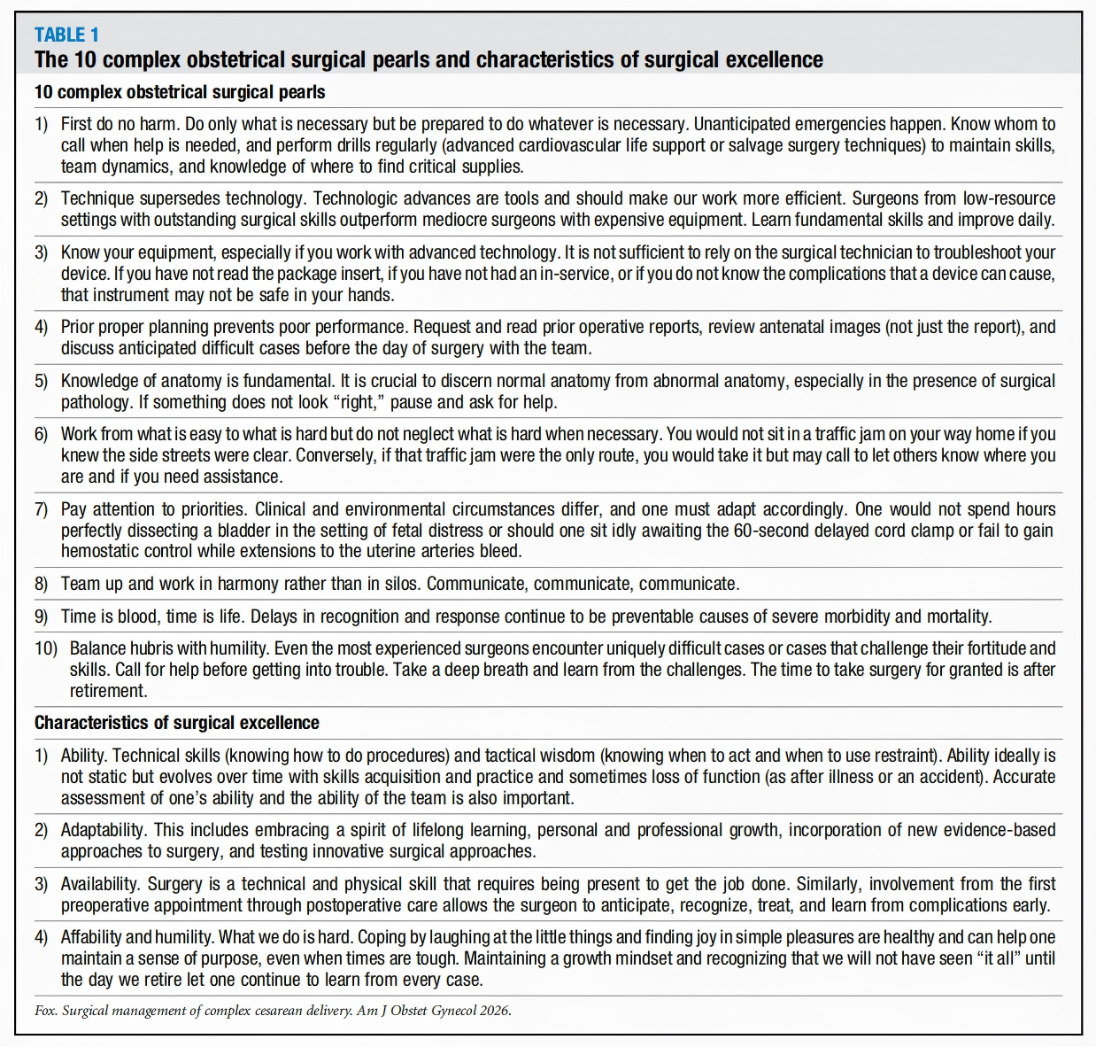
Table 1: Key Points of the Ten Complex Obstetric Surgery Truths and Traits of Surgical Excellence
These principles form the bedrock for managing the specific technical challenges described below.
Preoperative Preparation and Abdominal Incision Selection
Adequate preoperative preparation is the first step in managing a complex cesarean delivery.
This includes reviewing all available records, prenatal imaging (not just reports), and operative notes.
In non-emergent situations, updating the history and physical examination helps refine the surgical plan, secure additional resources, and approach challenges calmly.
The literature details the comparison of various abdominal incisions used for complex cesarean delivery (Table 2, Figure 1):
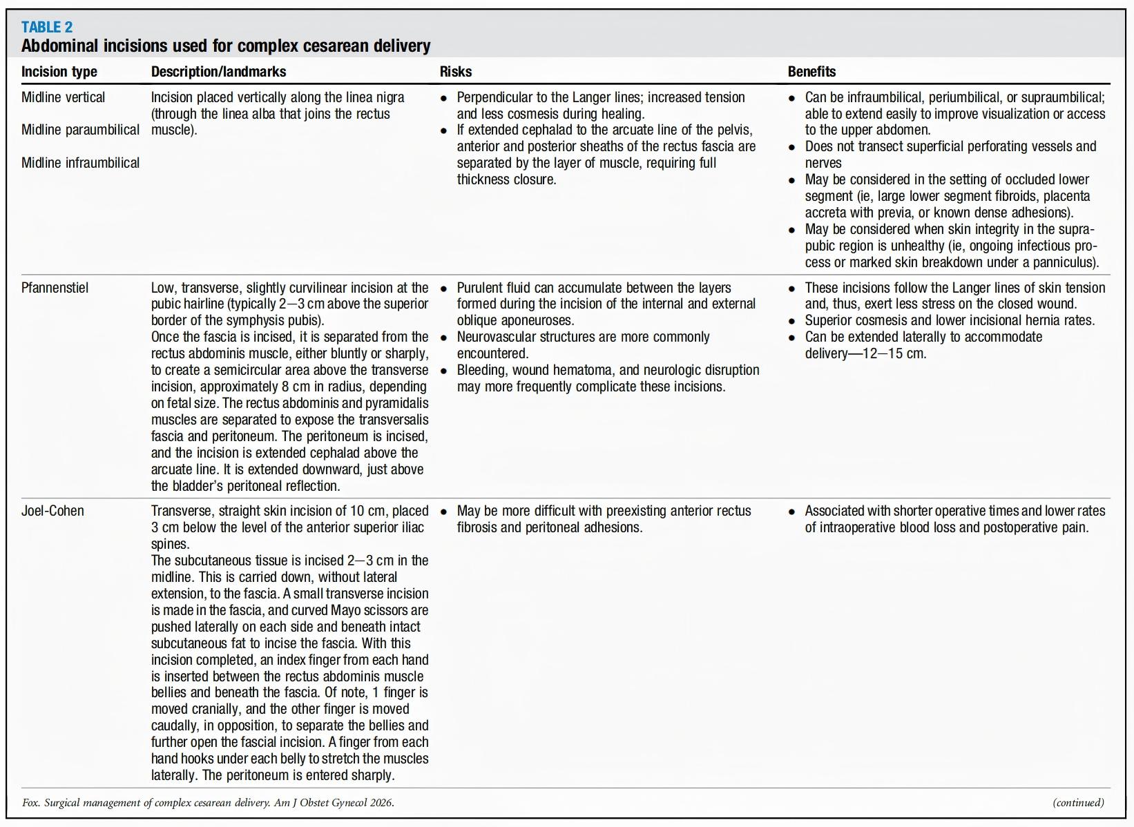
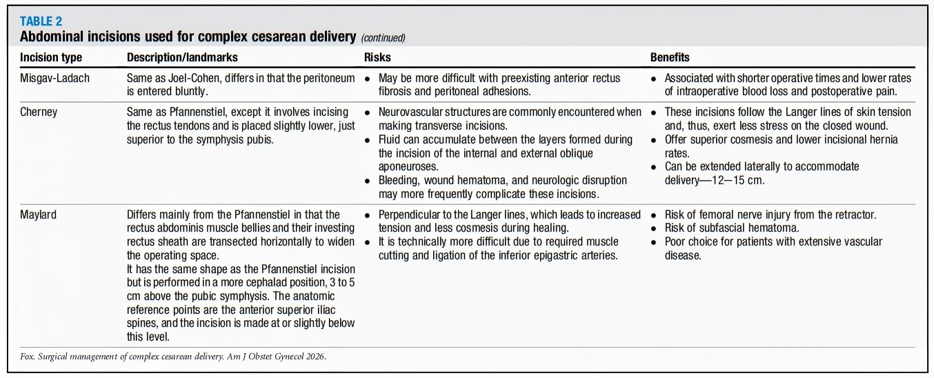
Table 2: Abdominal Incisions for Complex Cesarean Delivery
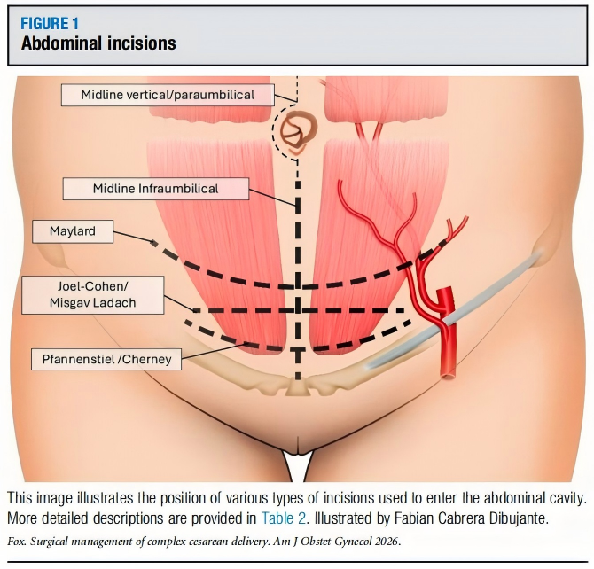
Figure 1: Abdominal Incisions
Midline Vertical Incision
A vertical incision along the linea alba.
Advantages: Easy to extend cephalad to improve visualization and access to the upper abdomen; does not transect superficial perforating vessels and nerves.
Useful for: Cases with a blocked lower uterine segment (e.g., large fibroids, placenta previa with accreta).
Disadvantages: Perpendicular to skin tension lines, resulting in greater tension and poorer cosmesis during healing; if extended cephalad beyond the arcuate line, requires full-thickness closure of the anterior and posterior rectus sheaths.
Pfannenstiel Incision
A low transverse curved incision 2-3 cm above the pubic symphysis.
Advantages: Follows skin tension lines, resulting in lower wound stress, better cosmesis, and lower incidence of incisional hernia; can be extended laterally to 12-15 cm.
Disadvantages: Pus can accumulate between the layers of the internal and external oblique aponeuroses; neurovascular structures are more frequently encountered, with slightly higher risks of bleeding, hematoma, and nerve injury.
Joel-Cohen Incision
A 10-cm transverse straight skin incision 3 cm below a line connecting the anterior superior iliac spines. Employs a specific technique for fascial incision and separation. Associated with shorter operative time and lower rates of intraoperative blood loss and postoperative pain.
Misgav-Ladach Incision
A modification of the Joel-Cohen incision emphasizing minimal tissue handling.
Maylard Incision
A transverse incision that transects the rectus muscles, used in extreme situations requiring extensive exposure.
Cherney Incision
An incision that detaches the rectus muscles from their insertion on the pubic symphysis, also used for situations requiring excellent pelvic exposure.
✿ Selection Principles
Incision choice should be based on: anticipated surgical complexity, patient body habitus, previous incisions, and surgeon experience.
For cases with known severe adhesions, large lower segment fibroids, or placenta accreta spectrum disorders with previa, a midline vertical incision may offer better exposure and extension flexibility.
Core Challenge One: Management of Adhesions
The incidence of adhesions after cesarean delivery is 46%-65% and increases with the number of cesarean sections. Adhesions remove normal tissue elasticity, distort anatomical planes, and increase the risk of injury to adjacent organs (bladder, bowel).
✿ General Management Principles
Avoid blunt, forceful dissection within adhesive tissue planes, which can lead to unpredictable tearing. Dissection should begin in the area that appears most normal, with the least fibrosis, carefully proceeding towards the most pathologically altered areas. The goal is to identify the correct tissue plane.
• Omental Adhesions: The omentum is highly vascular. (Figure 2)
During management, start at the narrowest point of the adhesion (often near the lateral abdominal wall or bladder). For dense adhesions, first create an avascular "window" within them, then serially clamp, divide, and ligate.
Key point: Always palpate behind the adhesion to avoid hidden bowel.
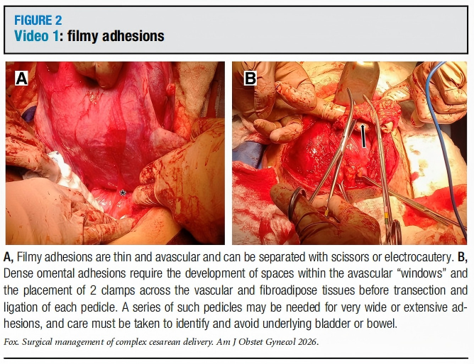
Figure 2: Membranous Adhesion
• Myometrial Adhesions: Dense, rubbery adhesive bands between the uterine myometrium and the rectus muscles. (Figure 3)
During management, first use a finger to delineate its borders and exclude bowel or omentum, then carefully divide with electrocautery, ensuring hemostasis of the myometrial edges. Bleeding from the base can be controlled with hemostatic agents or figure-of-eight sutures.
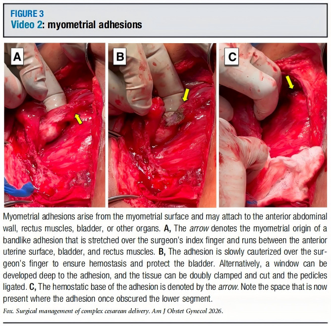
Figure 3: Myometrial Adhesion
• Bladder Adhesions: The bladder is the most commonly injured organ. Adhesions can cause it to be abnormally high-lying. (Figure 4)
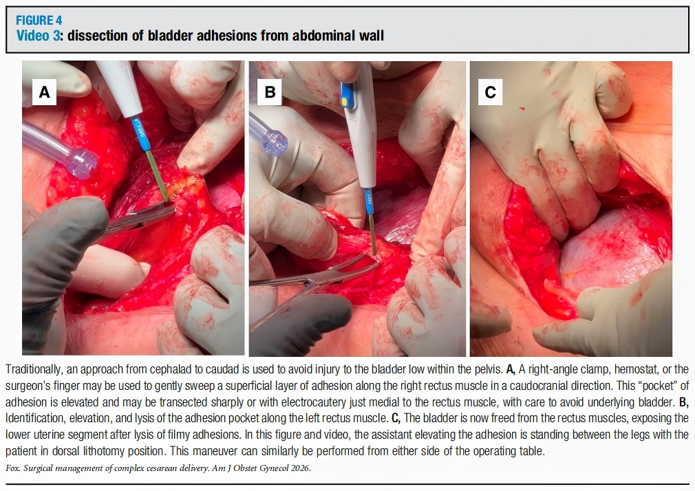
Figure 4: Separation of Vesicouterine Peritoneal Adhesion
◦ Approach: The traditional cephalad-to-caudad approach may be unsafe. The literature describes a caudad-to-cephalad approach: using a right-angle or hemostat clamp, carefully perform blunt and sharp dissection along the midline from caudad to cephalad, "lifting" the adherent rectus muscle off the bladder surface and dividing it.
◦ Pelosi Maneuver (Post-bladder Lower Uterine Segment Bypass): The peritoneum is incised medial to the round ligament, above the vesicouterine fold. A finger is inserted into the paravesical space, probing medially and inferiorly to assess if a safe separation plane exists between the bladder and the lower uterine segment. This helps evaluate the severity of adhesions. (Figure 5)
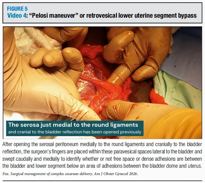
Figure 5: The "Pelosi Maneuver" or Post-bladder Lower Uterine Segment Bypass
◦ Bladder Dye Test: Clamping the urinary catheter and instilling sterile saline or diluted methylene blue into the bladder helps clarify bladder boundaries and detect occult injuries.
• Bowel Adhesions: Relatively uncommon. (Figure 6)
If they do not obstruct the surgical field, they can be left alone. If intervention is necessary, follow the "easy to hard" principle, gently pushing tissue aside with an instrument (e.g., the heel of a right-angle clamp) to find a plane of separation. Use fine scissors (e.g., Metzenbaum) for small, sharp movements. If bowel injury occurs, immediately isolate that segment and call for specialized assistance.
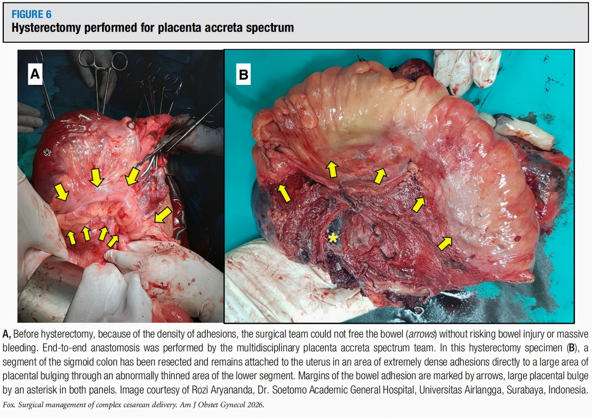
Figure 6: Hysterectomy for Placenta Accreta Spectrum
Core Challenge Two: Uterine Incision Extension, Dehiscence, and Rupture
• Uterine Incision Extension
Often extends laterally or caudally, increasing the risk of hemorrhage, hematoma, and ureteral injury. Common in preterm cesarean (narrow lower segment) or with a deeply engaged fetal head (thin lower segment).
◦ Management: The primary goals are hemostasis and adequate exposure. Initial control can be achieved with atraumatic clamps (e.g., ring forceps, Allis clamps) on the bleeding extended angles. Adequate bladder mobilization to expose the apex of the extension is essential. During repair, assess if the extension involves the posterior uterine wall and its proximity to the uterine vessels and ureters.

Figure 7: Repair of Uterine Incision Extension
• Uterine Dehiscence
Refers to thinning or separation of the uterine myometrium with an intact serosal layer (i.e., a "cesarean scar niche") following a previous cesarean or myomectomy.
◦ Management: The key is to place the uterine incision in healthy myometrium. Sometimes, an incision needs to be made more cephalad, above the dehiscence. For a well-defined focal dehiscence, consider excising the weak scar tissue and reapproximating: after adequate bladder mobilization, excise unhealthy tissue until normal, circumferential myometrial edges are obtained, then close in layers.

Figure 8: Uterine Dehiscence
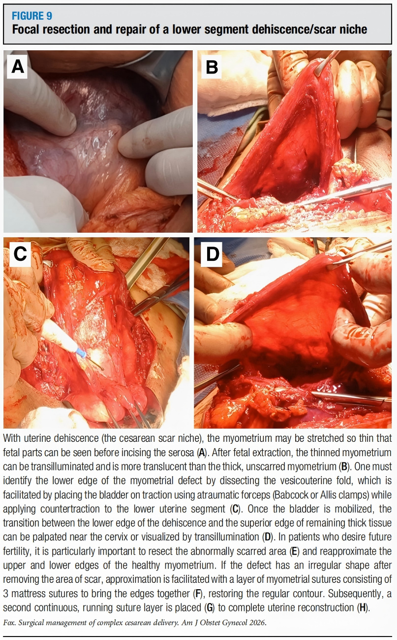
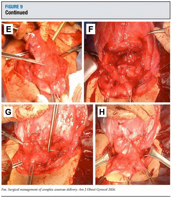
Figure 9: Focal Excision and Repair of Lower Segment Dehiscence/Scar Niche
• Uterine Rupture
A full-thickness tear at the site of a uterine scar, with the amniotic sac or fetal contents exposed to the abdominal cavity. It is an obstetric emergency with high risk of maternal hemorrhage and fetal hypoxia.
◦ Recognition:
▪ Maternal Signs: Include sudden severe abdominal pain (especially at the scar site), pain unrelieved by analgesia, signs of shock (hypotension, tachycardia).
▪ Fetal Signs: Include sudden fetal heart rate deterioration (recurrent decelerations, sinusoidal pattern, prolonged bradycardia), or even "recession" of the presenting part.
◦ Management:
The goal is rapid delivery of the fetus and control of maternal hemorrhage. Standard preoperative preparation steps may need to be omitted. Upon entering the abdomen, a new incision is often unnecessary; the fetus can be delivered through the rupture site. Immediate manual aortic compression or use of a uterine tourniquet. Carefully examine the anatomy and repair the rupture. If repair is impossible or hemorrhage uncontrollable, hysterectomy is required. Massive transfusion protocol must be activated.
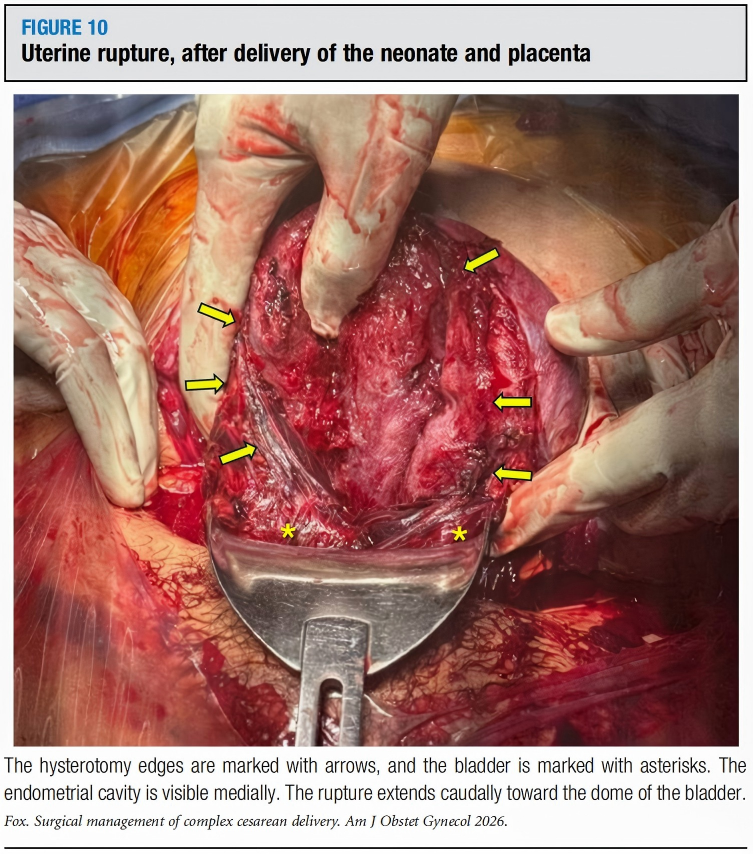
Figure 10: Uterine Rupture after Delivery of Newborn and Placenta
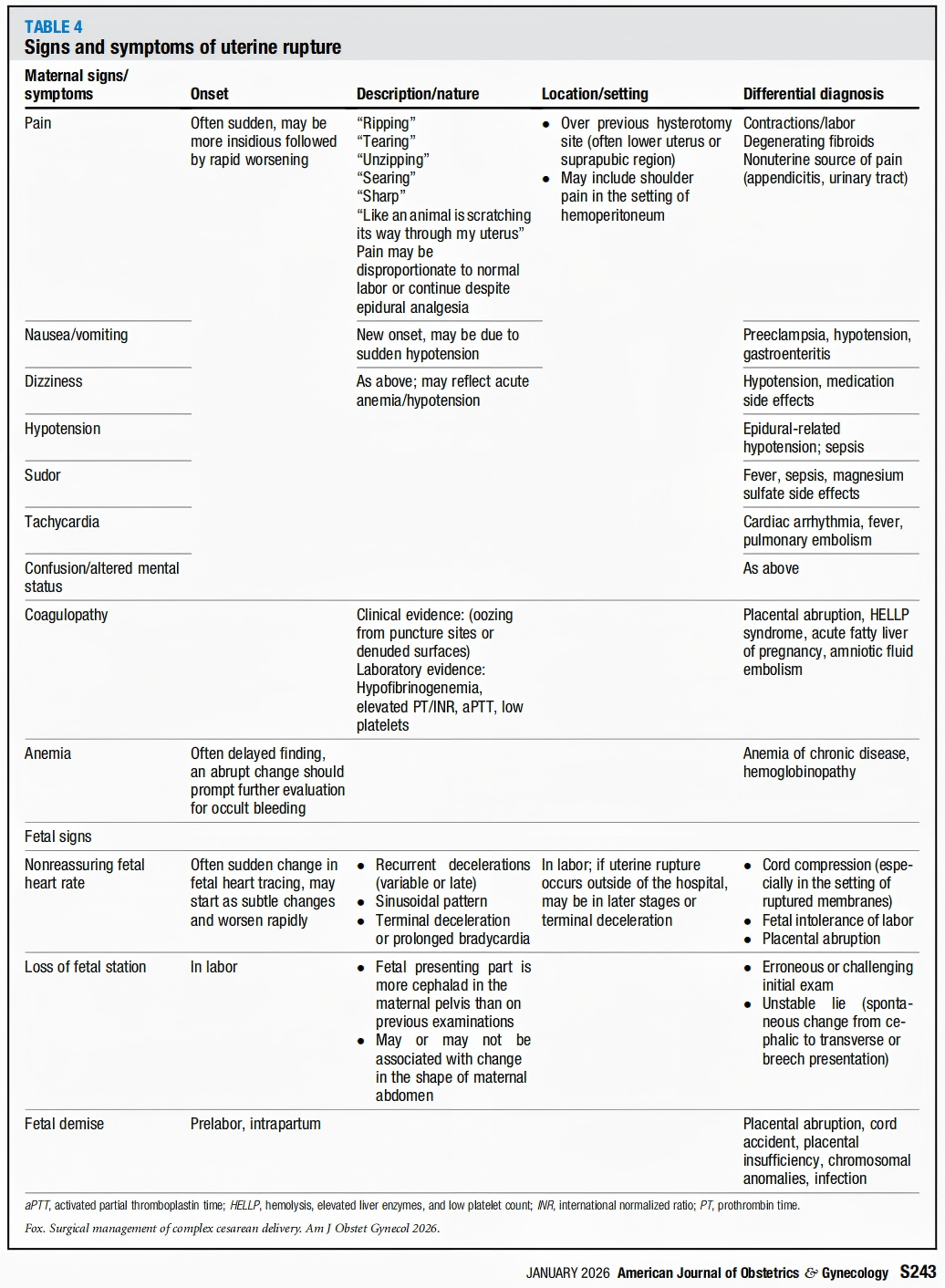
Table 4: Signs and Symptoms of Uterine Rupture
Core Challenge Three: Uterine Fibroids and Cesarean Myomectomy
Fibroids increase surgical complexity. The risk primarily depends on their location, size, and number. Large or multiple fibroids may obstruct uterine incision formation or closure, potentially necessitating a fundal or posterior uterine incision.
• Cesarean Myomectomy
Traditionally discouraged due to concerns about increased bleeding risk. However, recent observational studies and meta-analyses suggest that myomectomy concurrent with cesarean delivery may be safe and feasible in experienced hands and with proper preparation.
Studies show that while transfusion rates and estimated blood loss are increased compared to cesarean alone, postoperative hemoglobin changes and other complication rates are similar. Long-term benefits include relief of gynecologic symptoms and avoidance of a second surgery.
◦ Decision Points: Require careful assessment. Fibroids located laterally or near the cervix may significantly distort the course of the uterine vessels and ureters, substantially increasing surgical risk. If myomectomy is decided upon, it is recommended to incise the myometrium at the thinnest part of the fibroid and carefully enucleate it. Hemostatic adjuncts (e.g., antifibrinolytics, vasopressin, uterine tourniquet) can be used. Efforts should be made to avoid penetrating the endometrium to reduce the risk of future placenta accreta.
Core Challenge Four: Distorted Anatomy
1. Abnormally Positioned Vesicouterine Fold
In cases of a deeply engaged fetal head, large fibroids, or abnormally stretched lower segment, the vesicouterine fold is drawn relatively cephalad. (Figure 11)
◦ If unrecognized, placing the incision at the conventional distance can easily lead to bladder injury or an incision made on the cervix or even vagina.
◦ The key is to identify the anatomical attachment point of the vesicouterine fold on the uterus and use that as the guide for incision placement.
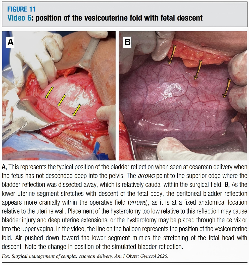
Figure 11: Changing Position of the Vesicouterine Fold with Fetal Head Descent
2. Uterine Incarceration and Sacculation
Incidence approximately 1/3000-1/10000.
◦ The gravid uterus (often retroverted) becomes trapped within the pelvis, unable to ascend into the abdomen.
◦ As pregnancy continues, the lower uterine segment becomes overstretched and thinned, forming a "sacculation."
3. Impact on Cesarean Delivery
Causes severe distortion of normal anatomical landmarks.
◦ The stretched bladder and lower segment are abnormally cephalad, and the cervix may also be displaced.
◦ Preoperative diagnosis and ultrasound mapping are crucial; the superior border of the bladder and the internal cervical os must be clearly identified to avoid injuring the displaced bladder, cervix, or posterior uterine wall during abdominal entry and hysterotomy.
4. Uterine Torsion
Rotation of the uterus along its longitudinal axis beyond 45 degrees. Rare, often associated with fibroids or uterine anomalies.
◦ On ultrasound, placental location may appear changed (anterior becomes posterior).
◦ During cesarean, an inadvertent posterior transverse incision may be made; after repair, careful inspection of the uterine vessels and ureters is needed.
Core Challenge Five: Management of Major Hemorrhage
Prevention is better than cure. Modern cesarean techniques aim to reduce unnecessary handling and protect small vessels.
• Basic Hemostatic Strategies
1. Hemostatic Approach (Figure 13)
In cases anticipated to have significant bleeding, carefully cauterize or ligate vessels in each layer of the abdominal wall.

Figure 13: Hemostatic Abdominal Approach
2. Uterine Incision Edge Clamping (Figure 14)
Using ring or Green-Armytage forceps to clamp the lateral edges of the uterine incision longitudinally can effectively compress bleeding venous sinuses (estimated bleeding rate 350-800 mL/minute). They can be advanced progressively during suturing.
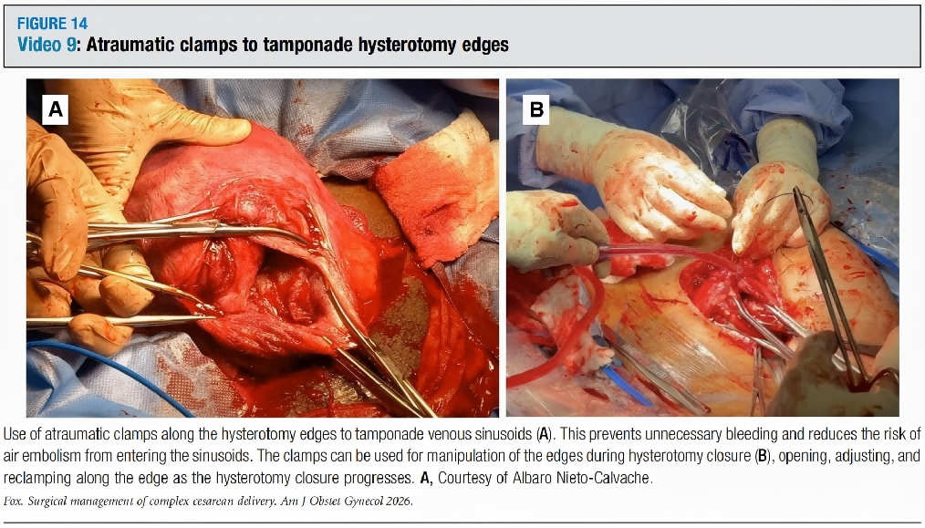
Figure 14: Atraumatic Clamps Used to Compress Uterine Incision Edges
3. Manual Compression
An assistant can use both hands to "sandwich" compress the lower uterine segment anteriorly and posteriorly, or compress the parametrium from the sides to temporarily control uterine artery flow.
4. Uterine Tourniquet (Figure 15)
Using a Penrose drain, sterile glove cuff, or Mersilene tape to encircle the lower uterine segment, temporarily compressing the uterine arteries and vaginal branches.

Figure 15: Uterine Tourniquet
5. Aortic Compression (Figure 16)
The most direct emergency measure. The surgeon or assistant directly compresses the abdominal aorta intra-abdominally with a hand, rapidly reducing perfusion pressure to the uterus and pelvis, buying time for definitive hemostasis.

Figure 16: Manual Aortic Compression
• Advanced Interventional Measures
In cases of refractory hemorrhage, consider aortic balloon occlusion (REBOA) or aortic clamping by a vascular surgeon, but this requires specialized team support.
Selection of Surgical Materials and Adjuncts (Table 3)

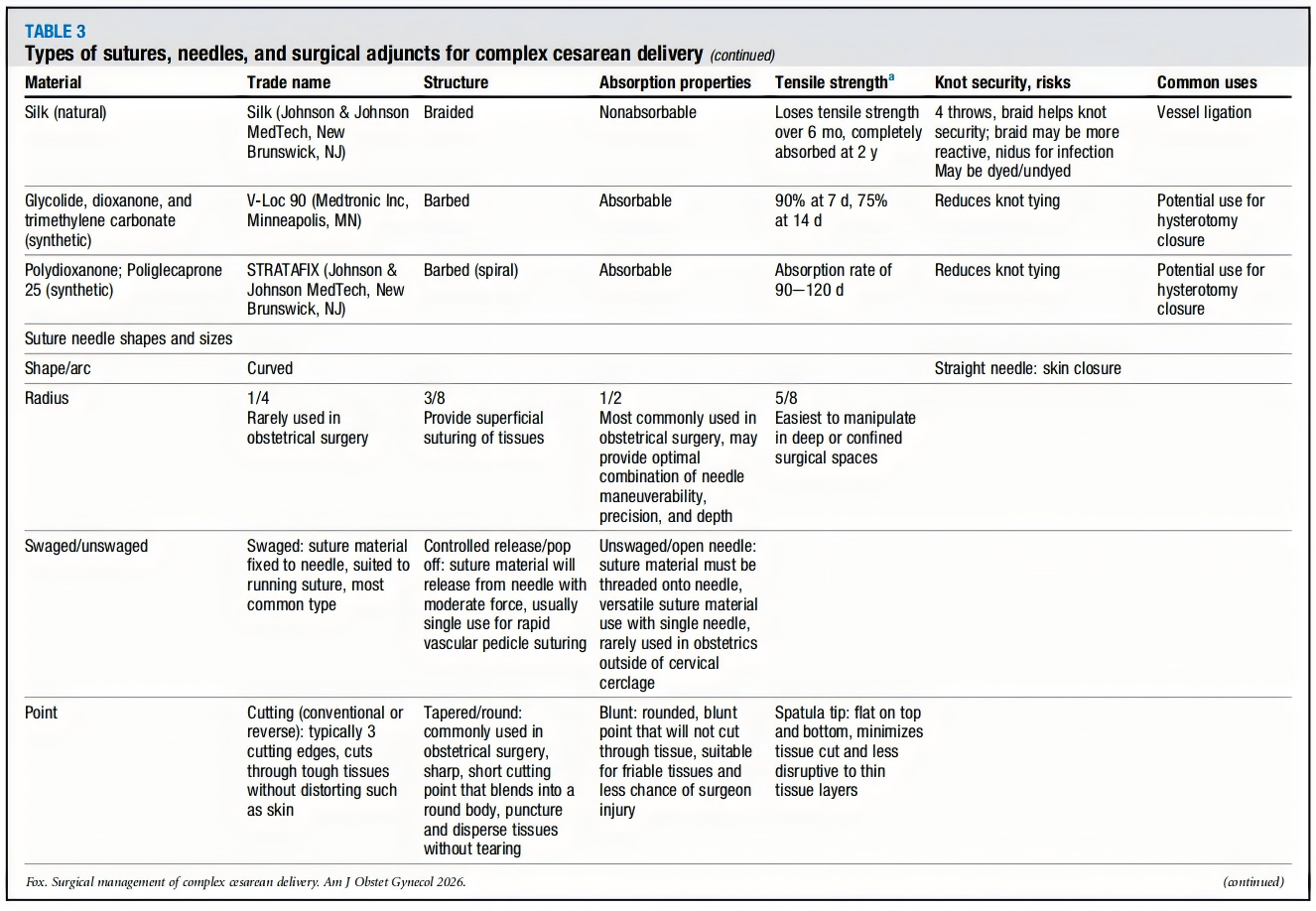

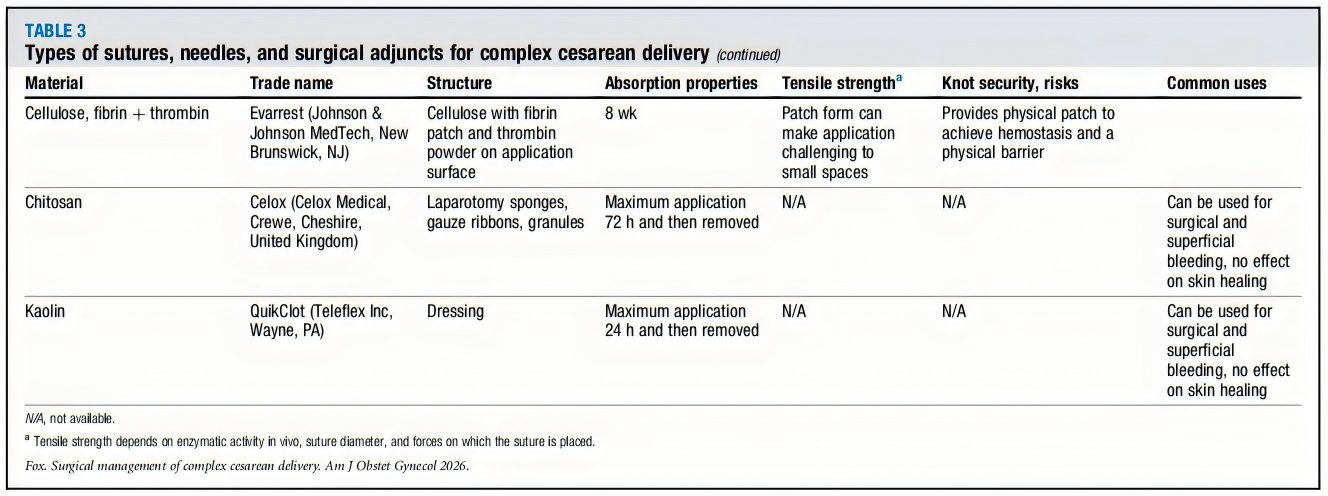
Table 3: Types of Sutures, Needles, and Surgical Adjuncts for Complex Cesarean Delivery
The literature provides a detailed review of sutures, needles, and hemostatic adjunct materials.
• Sutures: Selected based on tissue healing time and tensile strength requirements.
◦ For example: Fascial closure often uses Polydioxanone (PDS, slow-absorbing); myometrial closure often uses Polyglactin 910 (Vicryl, faster-absorbing) or Poliglecaprone 25 (Monocryl); subcutaneous closure can use fast-absorbing Polyglactin 910 (Vicryl Rapide). Barbed sutures (e.g., V-Loc, STRATAFIX) can reduce knot tying, and their potential for uterine incision closure is being explored.
• Needles: Curvature (1/2 circle most common), point (taper point most common, for delicate tissue like myometrium; cutting for skin), and size are chosen based on tissue depth and thickness.
• Hemostatic Agents: Include absorbable gelatin sponge (Gelfoam, provides tamponade), oxidized cellulose (Surgicel, various forms), thrombin preparations (Thrombin-JMI), thrombin-gelatin matrix (Floseal, suitable for irregular surfaces), fibrin sealant (Tisseel), etc.
◦ Understanding their properties, absorption time, and precautions (e.g., some not for skin, potential allergic reactions) is necessary.
Extreme Situation
• Resuscitative Cesarean Delivery: The literature mentions that cesarean delivery performed during cardiopulmonary resuscitation is one of the most technically and psychologically challenging surgical scenarios.
◦ The goal is to deliver the fetus within 4-5 minutes of maternal cardiac arrest to optimize maternal and neonatal survival.
◦ This demands extremely rapid decision-making and surgical execution, often requiring abandonment of standard sterile preparation steps.
Summary and Insights
This review systematically outlines modern management principles for complex cesarean delivery.
Its core message is: With rising cesarean rates and expanding indications, obstetricians must be prepared to manage complex scenarios like adhesions, fibroids, uterine dehiscence/rupture, and major hemorrhage. Success depends on:
• Solid anatomical knowledge and surgical fundamentals.
• Meticulous preoperative planning and multidisciplinary collaboration.
• Adaptability and clear priority judgment (time is life).
• Proficiency with various surgical approaches, techniques, and materials.
Faced with increasingly complex cases, continuous surgical education, simulation training, and teamwork are key to ensuring maternal safety.
This article and its accompanying video atlas (e.g., the Toronto Surgical Video Atlas) provide valuable resources for obstetricians to address these challenges (surgical videos can be watched by copying the video webpage addresses from the original text).
Translator's Introduction
Chart Source: AJOG
Editor: Lily