





















"Standardized Diagnosis Procedure Guide for Common Vaginitis (2025)" – A Comprehensive Manual for the Full Diagnostic Process from Medical History to Microscopy, Moving Beyond "Intuition-Based" Management
"Standardized Diagnosis Procedure Guide for Common Vaginitis (2025)"
Abstract: This guide aims to instruct medical institutions at all levels to standardize the precise diagnosis and treatment of vaginitis, ultimately achieving the important goals of reducing disease burden and improving women's reproductive health. To facilitate clinicians in quickly grasping core diagnostic and therapeutic points and standardizing vaginitis diagnosis, this article systematically summarizes the guide's content. It aims to enhance the standardization and precision of clinical diagnosis and treatment, thereby better realizing the guide's objective of reducing disease burden and improving women's reproductive health.
This guide utilizes the Grading of Recommendations Assessment, Development and Evaluation (GRADE) method to classify the quality of evidence, as shown in Table 1.
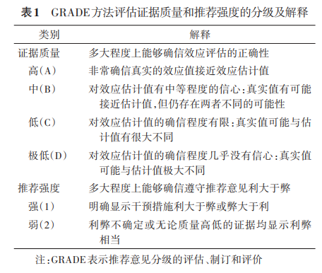
Vaginitis is a common gynecological disease, often caused by the invasion of exogenous pathogenic microorganisms or disturbances in the endogenous vaginal microbiota. Common clinical types include bacterial vaginosis (BV), aerobic vaginitis (AV), vulvovaginal candidiasis (VVC), trichomoniasis, and mixed vaginitis. To address prominent issues in the current diagnostic and treatment processes, such as inconsistent diagnostic criteria, significant variability in laboratory test results, and low identification rates of mixed infections, the National Clinical Research Center for Obstetrics and Gynecology Diseases, in collaboration with the National Center for Clinical Laboratories of the National Health Commission, has developed this guide. Based on GRADE methodology for evidence quality grading, referencing World Health Organization (WHO) guidelines and the latest evidence-based medical evidence, and integrating patient complaints, specialized physical examination findings, laboratory etiological tests, and vaginal microecological assessments, the guide establishes a multi-dimensional, standardized diagnostic process for different types of vaginitis. This process breaks through the limitations of traditional diagnosis relying solely on clinical symptoms or single laboratory indicators. By integrating a comprehensive diagnostic strategy combining clinical assessment and laboratory etiological examinations, it aims to comprehensively enhance the standardization and precision of vaginitis diagnosis in China, providing a reliable basis for effective clinical treatment.
Vaginitis is a collective term for vaginal diseases caused by infection, inflammation, or disruption of the normal vaginal flora. Its main manifestations include abnormal vaginal discharge, odor, itching, and vaginal discomfort. When the normal vaginal microecological balance is disturbed, abnormal discharge is easily produced. Contributing factors include exogenous pathogenic microorganism infections, endogenous flora imbalances, and other non-infectious factors. Clinical data shows that 70% to 90% of vaginal symptoms are caused by infectious factors. Common types mainly include bacterial vaginosis (BV), aerobic vaginitis (AV), vulvovaginal candidiasis (VVC, also known as vulvovaginal candidiasis), trichomoniasis, and mixed vaginitis. Global epidemiological surveys indicate that the incidence of vaginitis in women of reproductive age can reach 20% to 50%, with significant regional differences and seasonal characteristics. Incidence is generally higher in developing countries than in developed countries, and significantly increases during hot and humid seasons. This disease can not only cause symptoms such as vulvar itching and abnormal vaginal discharge but may also lead to serious complications like pelvic inflammatory disease (PID), infertility, premature birth, and significantly increase the risk of HIV and other sexually transmitted diseases (STDs).
In China's gynecology outpatient clinics, patients seeking treatment for vaginitis account for over 40% of visits. Current diagnosis and treatment of vaginitis face three core problems: first, clinical diagnostic criteria are not yet unified; second, laboratory testing technical levels are uneven, and the standardization of vaginal microecological assessment urgently needs improvement; third, lack of standardized infection diagnosis and treatment directly contributes to a vaginitis recurrence rate as high as 30% to 50%. To establish a systematic diagnostic framework for vaginitis, promote standardization of testing technology, improve the overall diagnosis and treatment level for vaginitis, and guide rational clinical medication, the National Clinical Research Center for Obstetrics and Gynecology Diseases, in collaboration with the National Center for Clinical Laboratories of the National Health Commission, organized leading experts nationwide in gynecology and clinical laboratory medicine to jointly formulate the "Standardized Diagnosis Procedure Guide for Common Vaginitis (2025)".
Gynecological History Taking Related to Vaginitis
Relevant gynecological history includes age, chief complaint, past medical history, menstrual and/or marital/obstetric history, and personal history.
01 Age Groups
Women can develop vaginitis throughout their entire life cycle, with common types showing significant differences across age groups.
1. Infancy and Early Childhood: The immune system is not fully developed, and sex hormone levels are low. Vaginitis at this stage is mostly caused by bacterial infections or external irritation.
2. Adolescence: The reproductive system matures gradually, with significant hormonal fluctuations. Increased estrogen levels and vaginal discharge make adolescent girls prone to bacterial vaginosis (BV), vulvovaginal candidiasis (VVC), and sexually transmitted pathogen-related vaginitis.
3. Reproductive Age: The reproductive system is mature, and hormonal changes are significant. High estrogen levels and increased vaginal discharge, coupled with factors like sexual activity, childbirth, contraceptive methods, and menstrual cycles, increase the risk of exogenous pathogen invasion and vaginal flora imbalance. This makes reproductive-age women susceptible to BV, aerobic vaginitis (AV), VVC, trichomoniasis, etc.
4. Pregnancy: Changes in estrogen and progesterone levels, altered local vaginal mucosal immunity, and increased cervical mucus and vaginal discharge elevate the risk of BV. Concurrent glucose metabolism and immune system abnormalities increase the incidence of VVC compared to non-pregnant states, with higher recurrence rates in the second and third trimesters.
5. Menopause: Postmenopausal women not receiving estrogen replacement therapy experience significantly reduced estrogen levels, leading to vaginal mucosal atrophy, dryness, and decreased resistance. This is often combined with urinary tract infections, making them prone to genitourinary syndrome of menopause (GSM) and BV.
02 Common Chief Complaints and Present Illness
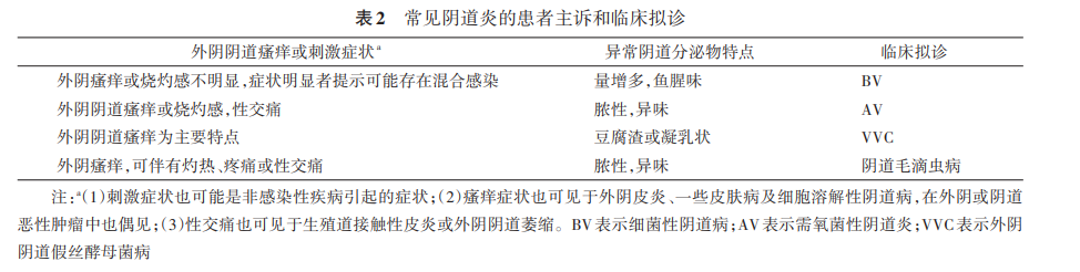
Patients with symptomatic vaginitis commonly present with: vulvar itching, vulvar irritation, which may or may not be accompanied by dysuria, urinary frequency, and dyspareunia; along with abnormalities in the amount, color, and odor of vaginal discharge. The degree of vulvar itching and characteristics of discharge abnormalities differ among vaginitis types. Clinicians can make preliminary judgments based on the patient's chief complaint and present illness (see Table 2).
03 Past Medical History
Relevant past medical history mainly includes history of vaginitis, medication history, history of diabetes mellitus, and history of sexually transmitted diseases (STDs), etc.
1. Medication History: Inquire about recent use of antibiotics, hormones, or other immunosuppressants, as these may affect the accuracy of laboratory test results.
2. Infection History: Record the frequency and specific types of vaginitis episodes in the past year to determine if it is a recurrent infection.
3. History of Diabetes Mellitus: Patients with poorly controlled blood glucose are more susceptible to vulvovaginal candidiasis (VVC).
4. STD History: Includes sexual activity, number of sexual partners, history of unprotected sex, and partner health status, used to assess high-risk factors related to vaginitis.
04 Menstrual and/or Marital/Obstetric History
Detailed information is needed regarding menstrual cycle, date of last menstrual period, sexual activity, marital/obstetric history, and the type and duration of contraceptive use. This information significantly impacts the timing of specimen collection and the choice of laboratory testing methods.
05 Personal History
Personal history mainly includes information on high-risk sexual behavior, adverse pregnancy history, history of vaginal foreign body or irritant contact, and adverse lifestyle habits.
1. High-risk Sexual Behavior: Mainly includes multiple sexual partners, unprotected sexual intercourse, sexual activity during menstruation, etc. Such behaviors increase the risk of vaginitis and STDs.
2. Adverse Pregnancy History: Women with a history of adverse pregnancy outcomes related to vaginitis require focused screening for genital tract infections during pregnancy to reduce the risk of recurrence.
3. Vaginal Foreign Bodies or Irritants: Includes long-term use of tampons, condoms, or use of irritating vaginal cleansing products, which may disrupt vaginal microecological balance and induce vaginitis.
4. Adverse Lifestyle Habits: Such as long-term smoking, alcohol abuse, frequent vaginal douching, etc., may all affect local vaginal immunity and flora balance, increasing the incidence of vaginitis.
[Recommendation 1]
A detailed patient history, including age, chief complaint, present illness, past medical history, menstrual and/or marital/obstetric history, and personal history, should be collected in the assessment of vaginitis to comprehensively evaluate infection risk and guide diagnosis (Recommendation Strength and Evidence Level: 1A).
Physical Examination Related to Vaginitis
The main pathological changes of vaginitis are located in the vagina. However, cervicitis and pelvic inflammatory disease (PID) can also present with increased vaginal discharge. Therefore, a comprehensive gynecological examination is required, including vulvovaginal, cervical, and pelvic examinations.
01 Vulvovaginal Examination
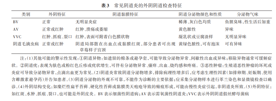
Vaginitis can present with corresponding changes in the vaginal mucosa and secretions. Different types of vaginitis exhibit differences in vulvar manifestations, characteristics of vaginal mucosal changes, and properties of vaginal discharge, as detailed in Table 3.
02 Cervical Examination
A cervical examination should be performed concurrently for patients with abnormal vaginal discharge. If cervical congestion, edema, friability, tendency for contact bleeding, and purulent discharge from the cervical canal are observed, cervicitis should be suspected.
➱ Differential Diagnosis: Cervicitis should be differentiated from cervical columnar ectopy, which is a normal physiological phenomenon where the columnar epithelium from the endocervical canal extends to the vaginal portion of the cervix. Furthermore, if infection with sexually transmitted pathogens like Neisseria gonorrhoeae or Chlamydia trachomatis cannot be ruled out, cervical secretions should be collected for relevant etiological testing.
03 Pelvic Examination
For patients with abnormal vaginal discharge, if signs such as cervical motion tenderness, uterine tenderness, and bilateral adnexal tenderness are present concurrently, pelvic inflammatory disease (PID) should be highly suspected. Further laboratory and imaging evaluations are recommended.
➱ Differential Diagnosis: Acute PID patients often present with systemic symptoms like fever (>38°C), chills, nausea, and vomiting. If an adnexal mass is palpated, it is necessary to differentiate between physiological follicles/benign cysts and malignant lesions like ovarian malignancies; the latter generally do not present with abnormal vaginal discharge, which can help distinguish them from infectious diseases.
04 Documentation of Examination
After history taking and physical examination, standardized documentation is required. Furthermore, when submitting laboratory test requests for vaginal discharge, key gynecological examination findings should be provided simultaneously to serve as a reference for laboratory diagnosis.
[Recommendation 2]
Physical examination should thoroughly evaluate areas such as the vulva, vagina, and cervix, noting mucosal changes and discharge characteristics, and document both positive and important negative signs (Recommendation Strength and Evidence Level: 1A).
[Recommendation 3]
A preliminary diagnosis of vaginitis can be made based on history and physical examination. To clarify the type of vaginitis and determine treatment, further laboratory etiological testing is needed (Recommendation Strength and Evidence Level: 1B).
Laboratory Examination of Vaginal Discharge
Different types of vaginitis can present with similar clinical manifestations and physical examination signs, and the same type of vaginitis can present with different physical findings depending on the severity and duration of the condition. Therefore, after completing history taking and physical examination, routine laboratory examination of vaginal discharge, especially etiological testing, is necessary. Initial assessment recommends the use of Gram-stained vaginal discharge smears and microscopic morphological examination. The entire testing process requires strict standardization, including correct specimen collection, transport, smear preparation, reading, and issuing of test reports (report results require dual review).
01 Preparation Before Sample Collection
1. Patient Preparation: Examination should preferably avoid the menstrual period. Instruct the patient to empty her bladder and assume the lithotomy position.
◦ Special Note: If the patient is menstruating, or if there has been sexual intercourse, sitz bath, vaginal douching, vaginal medication, or use of vaginal lubricants within 24 hours before sample collection, these may interfere with laboratory test results. Such conditions should be noted during sampling.
2. Sample Collection: Use a sterile swab to rotate and sample from the lateral vaginal wall where discharge is more abundant, ensuring the swab surface is clearly coated with discharge. Adequate and relatively consistent sampling volume should be ensured, avoiding insufficient or excessive sampling. After collection, place the swab in a sterile tube and transport it promptly for testing.
◦ After collection, affix a barcode for unique identification, including information such as patient name, age, hospital/outpatient number, physician's order, sample type, and test items.
3. Sample Transport and Reception: After collection, promptly label the sample with the collection time. Transport to the laboratory as soon as possible at room temperature, taking care to prevent tube breakage or leakage.
◦ Upon reception, verify sample information and collection time, and check if the sample quality meets testing requirements.
4. Handling of Unsuitable Samples: Unsuitable samples include, but are not limited to, unlabeled or mislabeled samples, container damage, and smears showing no nucleated cells on microscopy. The laboratory should register unsuitable samples, note the reason for unsuitability in the Laboratory Information System (LIS), and recommend clinical re-collection and submission.
02 Sample Examination
1. Testing Methods: Current routine laboratory methods for vaginitis diagnosis mainly include:
◦ Saline Wet Mount: Simple, rapid, and low-cost. Allows direct observation of trichomonads, fungal spores, and pseudohyphae. However, sensitivity is low, easily influenced by the examiner's experience, and cannot identify non-motile fungi.
◦ Fluorescent Staining: Uses special dyes for observation under a fluorescence microscope, significantly improving the detection rate and sensitivity for fungi and bacteria. However, equipment costs are higher, and operation is relatively complex.
◦ Dry Chemistry Enzymatic Methods (e.g., pH, hydrogen peroxide, leukocyte esterase): Offer standardization and semi-automation, with strong objectivity, suitable for large-scale screening. Can indicate flora imbalance but are indirect inferences, with possibilities of false positives or negatives, and cannot directly identify pathogen types.
◦ Molecular Biological Methods (e.g., PCR): High sensitivity and specificity for various pathogens (especially viruses, chlamydia, etc.). However, results are difficult to effectively distinguish between infection and colonization by opportunistic pathogens.
This guide establishes a standardized procedure for vaginal discharge morphological examination based on the Gram stain method, which is widely used in microbiology laboratories at all hospital levels. This method can simultaneously detect the four common vaginitis types (BV, AV, VVC, trichomoniasis) and mixed infections, while also assessing vaginal microecological status. It is suitable for routine implementation in hospitals at all levels, providing efficient and comprehensive etiological evidence for the clinical diagnosis of vaginitis.
2. Smear Preparation Requirements: Ensure even distribution of the sample on the slide, avoiding uneven thickness. Manual smear or automated liquid-based preparation methods can be chosen based on laboratory conditions. Smear preparation should be completed within 2 hours of sample reception.
◦ (1) Manual Smear Method: Keep the swab moist. Roll evenly in a single direction to create the smear, avoiding repeated smearing. Air-dry the smear naturally, then heat-fix.
◦ (2) Liquid-based Preparation Method: Use a specialized preservative solution for vaginal discharge. Follow the automated liquid-based cytology preparation process for centrifugation, preparation, fixation, and drying to ensure uniform, stable results.
3. Gram Staining Requirements: Manual Gram staining or automated stainers can be used. Use qualified staining reagents and strictly follow the staining procedure to ensure accurate bacterial staining and clear background.
◦ According to laboratory routine requirements and quality control frequency, use Gram-positive [Staphylococcus aureus (ATCC 25923)] and Gram-negative [Escherichia coli (ATCC 25922)] standard strains to perform quality control on staining reagents and the entire staining process.
4. Microscopic Examination: The core of vaginal microecological morphological examination is the identification and counting of formed elements in the smear, including epithelial cells, leukocytes, bacteria, fungi, and trichomonads. Standardize optical microscope observation parameters.
◦ (1) Manual Microscopy: For manual reading, an optical microscope typically uses 10x eyepieces. First, focus using a 10x objective, then use a 20x low-power objective to locate effective viewing fields. Subsequently, observe at least 20 fields under a 40x high-power objective to complete leukocyte and epithelial cell counts, and identify fungal spores, blastospores, pseudohyphae, and Trichomonas vaginalis. Finally, observe at least 20 fields under a 100x oil immersion objective to count bacteria with different staining and morphologies in the smear, taking the average for reporting.
◦ (2) Digital Microscope Scanning: Observation standards for digital scanning should align with manual microscopy field standards. Cell counting should follow the 40x high-power objective field standard, and bacterial counting should follow the 100x oil immersion objective field standard. Based on laboratory conditions, artificial intelligence-assisted analysis software with medical device registration qualifications can be used for auxiliary analysis. Before formally issuing clinical reports with this method, verification and confirmation via manual review are required to ensure result accuracy.
03 Morphological Report Content
The morphological report should focus on assessing flora density, flora diversity, dominant flora type, and changes in vaginal microbial morphological diagnostic indicators related to common vaginitis. Prioritize reporting morphological information with important clinical diagnostic value. All results should include normal reference ranges. Laboratories with the capability can provide image-text reports.
1. Vaginal Microecological Morphological Assessment Content
Includes flora density, flora diversity, and dominant flora type, each reported separately.
◦ (1) Flora Density Grading: Assess the total biomass of vaginal discharge flora by observing the average bacterial count per field under 10x eyepieces and 100x oil immersion, divided into 4 grades:
▪ Grade I: Average 1-9 bacteria per oil immersion field.
▪ Grade II: Average 10-99 bacteria per oil immersion field (normal reference range).
▪ Grade III: Average ≥100 bacteria per oil immersion field, with bacteria filling the field (normal reference range).
▪ Grade IV: Bacteria clump together, covering epithelial cells.
◦ (2) Flora Diversity Grading: Assess the number of bacterial morphological types in the smear, divided into 4 grades:
▪ Grade I: 1-3 bacterial morphologies.
▪ Grade II: 4-6 bacterial morphologies (normal reference range).
▪ Grade III: 7-9 bacterial morphologies (normal reference range).
▪ Grade IV: ≥10 bacterial morphologies.
◦ (3) Dominant Flora Type: In vaginal microecological assessment, the flora with the highest biomass or density is the dominant flora, which determines flora function. Report common dominant flora types based on Gram stain morphology:
▪ a. Gram-positive medium or large rods: Suggest Lactobacillus as the dominant flora, beneficial bacteria maintaining vaginal acidity and inhibiting pathogen proliferation.
▪ b. Gram-positive or variably stained short rods: Suggest Gardnerella as the dominant flora, possibly associated with bacterial vaginosis (BV).
▪ c. Gram-negative or variably stained curved rods: Suggest Mobiluncus as the dominant flora, possibly associated with BV.
▪ d. Gram-positive cocci: Suggest Streptococcus or Staphylococcus as dominant flora, possibly associated with aerobic vaginitis (AV).
▪ e. Gram-negative rods: Suggest Escherichia coli as the dominant flora, possibly associated with AV.
◦ Normal vaginal microecological morphological indicators are: Flora Density Grade II-III, Flora Diversity Grade II-III, Dominant Flora: Gram-positive medium or large rods.
2. Nugent Score for Diagnosing BV
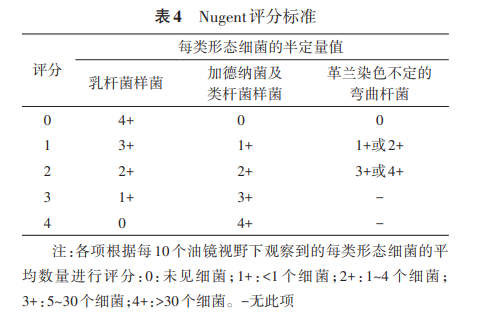
The Nugent score is currently the laboratory "gold standard" for diagnosing bacterial vaginosis (BV). After Gram staining the smear, observe under oil immersion. Score three bacterial morphological types according to the criteria in Table 4, sum the scores, and issue an etiological diagnostic report. Judgment criteria: 0-3 points = normal; 4-6 points = intermediate; ≥7 points = BV.
3. AV Score for Diagnosing AV
Currently, understanding of aerobic vaginitis (AV) is relatively recent, and no unified international diagnostic standard exists, with countries including the United States lacking clear diagnostic criteria. The 2018 European guidelines related to genital tract infections proposed laboratory diagnostic criteria for AV based on saline wet mount.
◦ Limitations of Saline Wet Mount:
▪ (1) Uses only unstained saline wet mount with a magnification of only 40x, insufficient for clear differentiation of bacterial morphology.
▪ (2) Bacteria are small under the microscope and can move with fluid, requiring phase-contrast microscopy for optimal observation.
▪ (3) Saline wet mounts cannot be stored long-term, preventing retrospective review.
◦ This guide recommends the AV diagnostic criteria proposed by Chinese scholars, combining Gram-stained microscopy with clinical features (such as vaginal mucosal congestion, yellow vaginal discharge), detailed in Table 5. The Gram stain method is the internationally accepted standard for laboratory bacterial morphological examination, providing more accurate results. Furthermore, stained smears can be stored long-term and reviewed, suitable for AV differential diagnosis and diagnosis of related mixed infections, while also facilitating the application of artificial intelligence-assisted analysis.
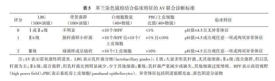
◦ Score each item combining morphology and clinical features. A total score ≥4 points can diagnose AV: 4-5 points = mild, 6-7 points = moderate, 8-10 points = severe AV.
4. Laboratory Diagnosis of Vulvovaginal Candidiasis (VVC)
Observe at least 20 fields under a 40x objective. If fungal blastospores or pseudohyphae are found, the etiological diagnostic report should state: "Fungal blastospores or pseudohyphae detected, clinically significant, requires comprehensive diagnosis combined with clinical symptoms." If not found, report: "No fungal blastospores and/or pseudohyphae found."
◦ For initial VVC diagnosis, or for follow-up/recurrent patients with poor empirical treatment response, if fungal blastospores or pseudohyphae are still detected, the sample should be sent for fungal culture, species identification, and in vitro antifungal susceptibility testing.
5. Laboratory Diagnosis of Trichomoniasis
Trichomonas vaginalis dies and disintegrates quickly after leaving the body. Specimens should be processed promptly. Observe under 10x or 20x objective; if trichomonad morphology is seen, switch to 40x for confirmation. Etiological diagnostic report: "Trichomonas found." Observe at least 20 fields under 40x; if no trichomonad morphology is seen, report: "Trichomonas not detected." During microscopy, note the presence of numerous leukocytes, which may be associated with trichomoniasis. If clinical suspicion of Trichomonas vaginalis infection is high, collect vaginal discharge in 0.5-1 ml saline, prepare a wet mount immediately (must be completed within 15 minutes), and observe for motile Trichomonas vaginalis to report results. If necessary, nucleic acid molecular detection methods can be used for confirmation.
6. Laboratory Diagnosis of Mixed Vaginitis
Mixed vaginitis is diagnosed using vaginal microecological examination methods. For the four common vaginitis types—bacterial vaginosis (BV), aerobic vaginitis (AV), vulvovaginal candidiasis (VVC), and trichomoniasis—if at least two pathogens are simultaneously present, or if diagnostic criteria for two or more vaginitis types are simultaneously met, mixed vaginitis can be diagnosed.
04 Quality Management
1. Quality Assurance
The laboratory should establish and strictly implement standardized operating procedures for vaginitis etiological examination. When test results are questionable, verification and re-examination should be performed.
The laboratory should establish a comprehensive process management system, standardizing the entire process from specimen preparation to report issuance. This achieves synchronous detection of AV, BV, VVC, trichomoniasis, and mixed vaginitis, enhances consistency and standardization of testing work, and ensures quality control at all stages.
2. Quality Control
◦ New batches/lots of Gram staining reagents require internal quality control (IQC) and comparison of specimen staining results with reagents in use before being approved for routine testing.
◦ Routine IQC can be performed weekly or daily alongside specimens. When uncertain test results occur, quality control should be initiated promptly to check reagents and staining procedures, ensuring Gram stain result accuracy and reliability.
◦ Regular proficiency testing and comparison for morphological examination personnel should be conducted and documented. Internal laboratory comparisons are recommended twice a year, with 10 specimens each time. Inter-laboratory comparisons should be conducted twice a year, with 5 specimens each time. Personnel assessment can use 50 microscopic images; inter-personnel agreement rate should be ≥90%, and each examiner's accuracy rate should be ≥90%.
◦ Additionally, the laboratory should conduct at least one human-machine comparison annually internally, with a false-negative rate ≤5%. At least one instrument comparison between laboratories should be conducted annually, with a result agreement rate >90%.
[Recommendation 4]
Laboratory testing for vaginal discharge should be requested and submitted as a routine and essential item for diagnosing vaginitis. The vaginal microecological Gram-stain morphological examination method is recommended. Testing should be performed in a standardized manner, encompassing the entire process management from sample collection, preparation, staining, microscopy, to reporting (Recommendation Strength and Evidence Level: 1A).
[Recommendation 5]
During the laboratory testing process, quality control should be performed regularly, especially using Gram-positive and Gram-negative standard strains for quality control during testing. Regular personnel and equipment comparisons should be conducted to ensure the accuracy and consistency of test results (Recommendation Strength and Evidence Level: 2A).
05 Diagnosis
Clinical diagnosis should be made by combining history, clinical symptoms and signs, and laboratory test reports. Patients diagnosed with vaginitis should subsequently receive targeted treatment based on the diagnostic conclusion. For patients whose diagnosis remains unclear or who experience symptom recurrence, microbial culture, identification, drug susceptibility testing, or nucleic acid testing is recommended. Following etiological confirmation, individualized precise treatment should be administered. Laboratories should regularly conduct multidisciplinary discussions and summaries of difficult cases with clinical departments to promote continuous improvement in diagnosis and treatment.
Summary
This guide systematically outlines the standardized diagnostic procedures for common vaginitis in obstetrics and gynecology in China, covering bacterial vaginosis (BV), aerobic vaginitis (AV), vulvovaginal candidiasis (VVC), trichomoniasis, and mixed vaginitis, including clinical assessment and laboratory examination standards. The GRADE method is used to grade evidence quality and recommendation strength, emphasizing the synergistic role of history taking, physical examination, and laboratory etiological testing (such as Gram-stain morphological examination) to ensure diagnostic precision. Simultaneously, it standardizes the entire process management from sample collection, quality control, to report interpretation, enhancing consistency and reliability in clinical practice. Furthermore, it emphasizes multidisciplinary team collaboration and individualized treatment, providing a scientific basis for the standardized diagnosis and treatment of vaginitis.
Source: National Clinical Research Center for Obstetrics and Gynecology Diseases, National Center for Clinical Laboratories of the National Health Commission. Standardized Diagnosis Procedure Guide for Common Vaginitis (2025) [J]. Chinese Journal of Obstetrics and Gynecology, 2026, 61(02): 89-96. DOI:10.3760/cma.j.cn112141-20250814-00378
Editor: Lily