





















Persistent infection with high-risk human papillomavirus (HR-HPV) is the primary cause of cervical precancerous lesions and cervical cancer. Cervical squamous intraepithelial lesions are classified as low-grade squamous intraepithelial lesions (LSIL, equivalent to CIN 1) and high-grade squamous intraepithelial lesions (HSIL, encompassing CIN 2 and CIN 3). Traditionally, CIN 2 diagnosed by cervical biopsy has been considered an indication for excisional treatment. However, multiple studies over the past decade have shown a high rate of spontaneous regression for CIN 2 within 24 months, especially in women under 30, whose risk of progression to invasive cancer is less than 0.5%. Nonetheless, the long-term risk of progression to invasive cancer rises again with prolonged conservative follow-up. Therefore, the core of clinical management for CIN 2 lies in achieving precise stratification, clarifying indications for conservative follow-up versus treatment, establishing monitoring/follow-up strategies, and formulating management plans for special populations. This has become an urgent clinical issue. The Colposcopy and Cervical Pathology Division of the Chinese Society of Birth Health and the Gynecological Oncology Branch of the Chinese Medical Association jointly organized experts to develop this consensus based on evidence-based medicine principles and the specific context of China. The consensus aims to guide the individualized and refined standardized management of CIN 2 in clinical practice.
I. Epidemiology and Natural History of CIN 2
1. Incidence of CIN 2
Globally, there is no uniform registration or statistical data for CIN 2 incidence. A UK cervical cancer screening study involving women not vaccinated against HPV indicated that approximately 2%-3% of women are diagnosed with CIN 2/CIN 3 annually, with the highest incidence (up to 10%) in the 25-29 age group. In China, the age-standardized incidence rates for histologically confirmed CIN 2 and CIN 3+ lesions are 1.3% and 1.2%, respectively.
2. Natural Regression Rate of CIN 2
Multiple international studies report that 50%-60% of CIN 2 lesions spontaneously regress to CIN 1 or normal cervical tissue within 2 years of follow-up, 23%-32% persist, and 18%-23% progress. Among these, the regression rate for women under 30 can reach 60%. Chinese studies show that among CIN 2 patients of reproductive age followed for 3-66 months, regression occurred in 63%-78.6%, persistence in 9.2%-22%, and progression to CIN 3 in 12.2%-15%. No progression to cervical cancer was observed in any of the study subjects.
II. Colposcopic and Histopathological Features of CIN 2
1. Colposcopic Features of CIN 2
Colposcopic diagnosis of CIN 2 has a relatively low concordance rate with histological diagnosis. Its features are intermediate between CIN 1 and CIN 3, as follows:
① Location: Most lesions are located within the cervical transformation zone, adjacent to the squamocolumnar junction (SCJ). They may appear as isolated lesions (Figure 1A) or be intermingled within larger lesions (Figure 1B).
② Color: After acetic acid application, the acetowhite change is less translucent and more durable than in CIN 1, but less dense and less persistent than in CIN 3.
③ Contour: Lesions often appear as patchy and mildly thickened. Contour alone is not sufficient to accurately differentiate CIN 2 from CIN 1 or CIN 3.
④ Margins: The border often lacks the typical irregular or geographic appearance characteristic of CIN 1.
⑤ Vascular Patterns: If present, vascular patterns are usually fine to medium in caliber, less prominent than in CIN 3. Intercapillary spacing is similar to or slightly larger than in CIN 1, and in rare instances approaches that seen in CIN 3 (Figure 1C). The presence of atypical vessels should raise suspicion for coexisting invasive cervical cancer with a high-grade lesion.
⑥ Iodine Staining: Lesions are usually iodine-negative (Figure 1D), though a partially iodine-stained yellowish-brown appearance may occasionally occur.
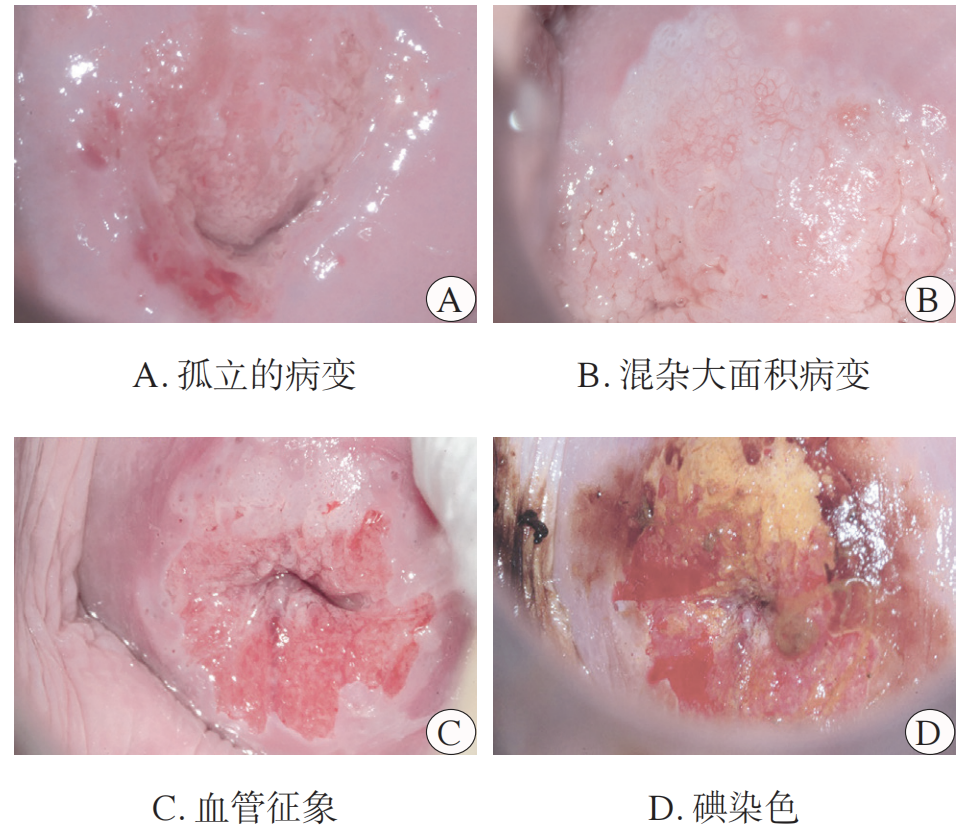
Figure 1: Colposcopic Images of CIN 2
Particular attention should be paid to thin HSIL (histological epithelial thickness <10 cell layers). On colposcopy, these lesions are often small foci, with 91.6% easily judged as low-grade impression, and 83.3% are histologically CIN 2. Therefore, during colposcopic biopsy, multiple biopsies should be taken from acetowhite areas, regardless of whether the colposcopic impression is metaplasia or a higher-grade abnormality.
Studies on endocervical curettage (ECC) show: in women with low-grade colposcopic impression and a Type 3 transformation zone, ECC did not detect CIN 2+ lesions in those with low-grade cytology [ASC-US/LSIL]. High-grade cytology [ASC-H or worse] was the only independent factor for ECC detection of CIN 2+. ECC provided a diagnostic gain of 2.9% for detecting CIN 2+ missed by colposcopic biopsy. Multivariate analysis suggests HPV 16 positivity is independently associated with additional diagnostic benefit from ECC. Currently, there is insufficient evidence to recommend routine ECC for all CIN 2 patients. However, it should be considered and accurately evaluated in cases with a Type 3 transformation zone, lesions extending into the endocervical canal, high-grade cytology, and persistent HPV 16/18 infection.
2. Histopathological Features of CIN 2
The pathological morphology of CIN 2 is intermediate between CIN 1 and CIN 3, sharing some features of both, leading to relatively low diagnostic agreement among pathologists. Its core features include squamous epithelial cell atypia, increased nuclear-cytoplasmic (N/C) ratio, presence of mitotic figures, and occasionally pathological mitotic figures. Atypical cells are mostly distributed in the lower half of the epithelium. The key distinction from CIN 3 is that CIN 2 retains some differentiation features, particularly the presence of intermediate and superficial squamous cells in the mid-to-upper layers. Koliocytes associated with HPV infection are often seen in the mid-superficial layers, similar to CIN 1. However, cellular proliferative activity and atypia are greater than in CIN 1, evidenced by higher N/C ratio in basal/parabasal layers, more frequent mitotic figures, which may also appear in the middle epithelial layers (Figure 2A, B). Additionally, CIN 2 must be differentiated from non-HPV-related epithelial proliferations and metaplastic lesions, such as immature squamous metaplasia, transitional cell metaplasia, and inflammatory reactive hyperplasia. Immunohistochemistry (IHC) has auxiliary value: the vast majority of CIN 2 show block-positive p16 staining, while only a minority of HPV-independent HSIL may be p16 negative. It should be noted that 20%-40% of CIN 1 can also be p16 positive; therefore, a diagnosis of CIN 2 should not be based solely on p16 positivity but requires integration with morphological features and HPV status. Regarding Ki-67 proliferation index, positive cells in CIN 2 can extend to the mid-upper epithelial layers, with a significantly higher proportion of positive cells than in CIN 1, though there is no unified diagnostic threshold (Figure 2C, D). Current evidence suggests that these IHC markers have no clear predictive value for CIN 2 regression or progression; clinical management decisions should still be based on a comprehensive assessment of clinical presentation, patient age, and other factors.
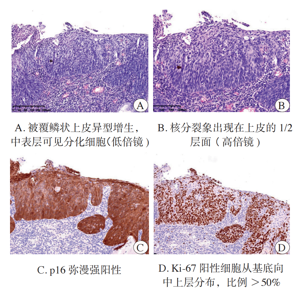
Figure 2: HE and IHC Staining of CIN 2
Pathological diagnosis of CIN 2 should be described systematically: specify the specimen type (biopsy or cone), and the diagnosis should be stated as High-Grade Squamous Intraepithelial Lesion/Cervical Intraepithelial Neoplasia Grade 2 (HSIL/CIN 2). p16 and Ki-67 IHC staining should be performed when necessary, describing the p16 staining pattern, Ki-67 positivity percentage, and the location of positive signals.
III. Conservative Management (Follow-up) for CIN 2
For patients diagnosed with CIN 2 by colposcopy-directed biopsy, it is crucial to accurately distinguish between two groups: a high-risk group, where there is a possibility of missed CIN 3+ or a high risk of CIN 2 persistence/progression to CIN 3+, who may benefit from diagnostic and therapeutic excisional treatment; and a low-risk group, with lower risks, who may be suitable for conservative follow-up with close monitoring and dynamic assessment, aiming to avoid immediate invasive treatment. Relevant factors for this assessment include patient age, fertility desire, cervical cytology results, HPV genotype, colposcopic visibility of the squamocolumnar junction (SCJ), lesion characteristics, and immune status.
(I) Key Determinants for Conservative Management of CIN 2
1. Age: Age is a core factor predicting CIN 2 regression or progression. Younger women, especially those <25 years, are in a sexually active phase with relatively high HPV infection rates, but their immune systems are more active and responsive, leading to mostly transient HPV infections. Studies show 88% regression of CIN 2 in women <25, with a mean regression time of 21 months. Data from China also indicate a significantly higher HSIL regression rate and shorter regression time (12 vs. 15 months) in those <25 compared to those ≥25. Women ≥30 years have a higher long-term risk of progression to invasive cancer; if conservative management is chosen for this group, more intensive monitoring and assessment are required.
2. HR-HPV Genotype: Studies show the progression risk for CIN 2 is significantly higher in HR-HPV positive individuals (25%) than negative (3%). HPV 16 carries the highest progression risk (71%), followed by HPV 18 (48%). CIN 2 patients with HPV 16 positivity showed 47% progression to CIN 3+ after 24 months of conservative follow-up. Multivariate analysis indicates HPV 16 positivity increases progression risk by 1.97-4.8 times.
3. Cytological Abnormality: CIN 2 patients with high-grade cytology (ASC-H or worse) have a 25%-60% risk of progression. Among them, those with HSIL cytology have a 3.8-5.0 times higher risk of colposcopy-missed CIN 3+, persistence, or progression. Furthermore, the time to regression for CIN 2 patients with HSIL cytology or HPV 16 positivity is 2-3 times longer than for those with low-grade cytology (ASC-US/LSIL) or non-HPV 16 positivity. However, no cervical cancer was detected during a median follow-up of 25 months in all patients.
4. Colposcopic SCJ Visibility: Whether the SCJ is fully visible on colposcopy is a key factor in conservative management decisions. Studies show the risk of missed cervical cancer is 0.6% vs. 0.3% (P=0.011) when the SCJ is not visible vs. visible. Chinese data also indicate the risk of missed lesions in CIN 2 patients increases 9.6-fold when the SCJ is not visible.
5. CIN 2 Lesion Characteristics: A domestic study showed CIN 2 lesions involving multiple quadrants had a lower regression rate (42%) and a higher progression rate (27% vs. 6%) compared to those involving a single quadrant (80% regression). Foreign studies also indicate lesion extent is a significant factor affecting regression at 3-month follow-up, with lesions confined to one quadrant regressing significantly more often than multi-quadrant lesions. Additionally, widespread involvement of endocervical crypts by high-grade lesions, presenting as expansile CIN, may increase progression risk, requiring careful assessment in clinical decision-making.
➱ Recommendations
① Women with CIN 2 aged <25 years: Conservative follow-up is the preferred recommendation, with treatment as an acceptable alternative.
② Women with CIN 2 aged ≥25 years with fertility desires (if concerns about the potential impact of excisional treatment on future pregnancy outweigh concerns about progression risk), may opt for conservative follow-up provided the SCJ and lesion upper border are fully visible, no CIN 2 is detected in the endocervical canal, there is no history of cervical lesion treatment, and conditions for standardized follow-up are met.
③ Conservative follow-up should be chosen with extra caution for those with high-risk features for persistence/progression, such as extensive lesions (>2 quadrants), expansile CIN, persistent HPV 16/18 positivity, or high-grade cytology (ASC-H, AGC, HSIL).
(II) Conservative Management Process and Monitoring for CIN 2
For CIN 2 patients planned for conservative management, a comprehensive assessment of medical history, cytology, colposcopic impression, and biopsy pathology is first required to exclude glandular lesions and suspicious invasive cancer. Patients must be fully informed about the goals, limitations of colposcopy (e.g., missed lesion risk), possibilities of persistence/progression, risks of delayed treatment, and the absolute necessity for strict adherence to follow-up schedules. Informed consent should be obtained. The specific process is detailed in Figure 3.
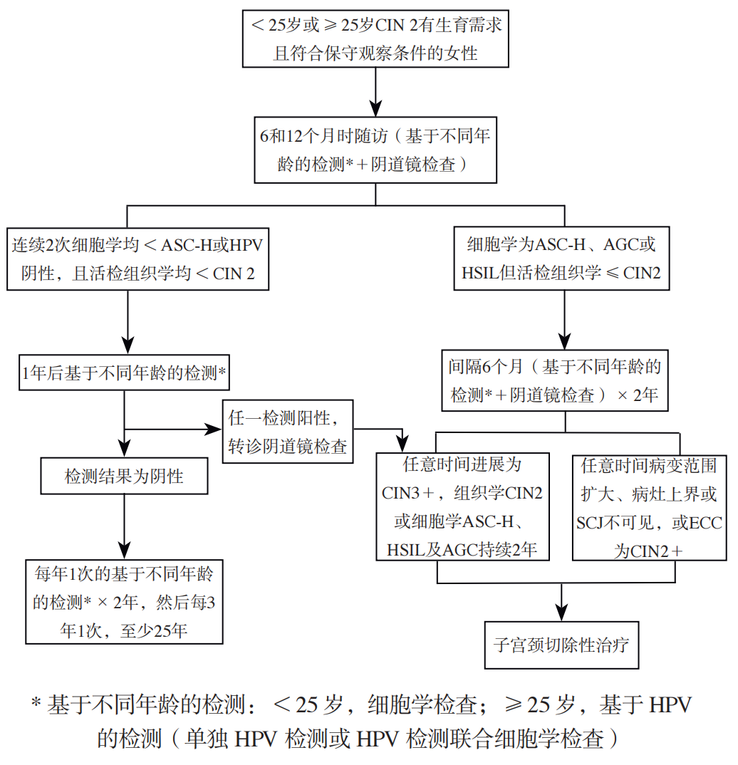
Figure 3: Conservative Management Process for CIN 2
1. Routine Monitoring for Conservative Management: Monitoring should be stratified by age. For those <25, cytology is recommended. For those ≥25, HPV-based testing (HPV test alone or co-testing with cytology) combined with colposcopic assessment is recommended. Directed biopsies ± ECC should be performed on all discontinuous acetowhite areas on colposcopy (regardless of whether the impression is metaplasia or higher-grade abnormality). While cervical biopsy may have some effect on promoting lesion regression, the risk of CIN 2 progression necessitates persistent dynamic monitoring.
2. Novel Monitoring Markers for Conservative Management: ① Methylation Testing: Studies show positive methylation testing combined with high-grade cytology can predict CIN 2 progression. Clinical regression rates were significantly higher in those negative for FAM19A4/miR124-2 methylation (74.7%) than positive (51.4%). ② HR-HPV Genomic Integration: Research indicates the risk of progression to HSIL is 5.6 times higher in the HR-HPV integration-positive group. An ongoing study on HR-HPV integration and CIN 2/3 natural history shows persistence/progression rates of 58.82% in the integration-positive group vs. 15.29% in the negative group. These markers are currently in the clinical research stage and require more large-scale study data for validation.
3. Indications for Terminating Conservative Management: ① If CIN 3+ is detected, or if CIN 2 persists for 2 years, cervical excisional treatment should be performed. ② Conservative management should be terminated if the lesion expands, extends into the endocervical canal, or if the upper border of the lesion/SCJ is not visible on colposcopy.
4. Follow-up Protocol After CIN 2 Regression: After two consecutive tests (6 months apart) show cytology <ASC-H, or HPV negativity with biopsy pathology <CIN 2, follow-up should be annual based on age stratification. If results remain negative, continue annual follow-up for 2 more years, then switch to 3-yearly follow-up. Total follow-up duration should be at least 25 years.
IV. Treatment for CIN 2
Primary treatment modalities for CIN 2 are divided into two categories: ① Conservative (Ablative) Treatments: including ablation (laser, cryotherapy, thermal coagulation, electrocautery, etc.) and photodynamic therapy (PDT). ② Cervical Excisional Treatments: primarily loop electrosurgical excision procedure (LEEP) and cold knife conization (CKC).
(I) Ablative Treatment
Ablative treatments are simple, require no or only local anesthesia, and have quick recovery, but do not provide a histological specimen for pathological assessment. Studies show no significant difference in clinical efficacy or pregnancy outcomes between CO₂ laser ablation and LEEP. Cervical cryotherapy and thermal coagulation are also effective for CIN 2. A systematic review showed patients with HSIL had a significantly higher risk of persistent/recurrent disease after ablation (cryo, thermo, laser) compared to excisional treatment (RR=1.65, 95%CI 1.25-2.19; RR=1.78, 95%CI 1.27-2.51 at 12 months), with comparable safety. Therefore, ablation should be chosen cautiously only after thoroughly excluding invasive cancer and glandular lesions.
Indications for Ablative Treatment: ① SCJ and entire lesion are fully visible. ② No histologically confirmed high-grade intraepithelial lesion in the endocervical canal. ③ Entire lesion is within treatable area. Invasive cancer must be ruled out before treatment. Ablation is not suitable if the lesion involves >75% of cervical surface area, extends into the canal, is associated with glandular lesions, there is a history of HSIL treatment, or during pregnancy or acute inflammation.
Precautions for Ablative Treatment: ① Reassess history, cytology, colposcopy, histopathology, and indications; obtain informed consent. ② Perform post-menses; avoid intercourse, douching, and vaginal medication for 3 days prior. ③ Local cervical anesthesia (intracervical or paracervical block) can be used. ④ Monitor patient response; transient vagal symptoms (flushing, dizziness, headache) may occur; mild lower abdominal pain is common and usually resolves spontaneously. ⑤ Postoperative vaginal discharge of varying degrees may last 10-20 days; fever, significant bleeding, or severe pelvic pain require prompt reevaluation. Avoid intercourse, swimming, and tub baths for 1 month post-treatment. Routine antibiotics are not needed; consider prophylaxis for those with prior PID. Regular follow-up is required.
(II) Photodynamic Therapy (PDT)
PDT is a targeted treatment combining a photosensitizer, specific wavelength light, and tissue oxygen. Its main advantage is minimal structural damage to the cervix and lesser impact on adverse pregnancy outcomes like miscarriage/preterm birth, making it suitable for young women with fertility desires.
Based on administration route and photosensitizer, common clinical PDT types include: ① 5-aminolevulinic acid PDT (ALA-PDT): A Chinese prospective cohort study showed a 92% regression rate at 6 months for CIN 2 treated with ALA-PDT. ② Hematoporphyrin derivative PDT (HpD-PDT): A small Chinese study showed 100% regression for CIN 2 and 84.2% for CIN 3 at 12-month follow-up post-HpD-PDT, but strict light avoidance is required post-treatment. ③ Hexaminolevulinate PDT (HAL-PDT): Uses an integrated device with photosensitizer and intravaginal cold light source. A global multicenter randomized double-blind placebo-controlled study showed 95% regression for CIN 2 after 1-2 HAL-PDT treatments.
Indications for PDT: Young women with fertility desires; fully visible SCJ and lesion upper border; no endocervical HSIL; no invasive cancer or glandular lesions. Caution is needed for extensive lesions. Use PDT protocols recognized by domestic/foreign authorities and validated in clinical trials for CIN 2.
Contraindications for PDT: Cytological/histological suggestion of glandular lesions, invasive cancer, or high clinical suspicion of malignancy; allergy to red light or related light sources; porphyria or allergy to ALA, gel, or components; pregnancy and lactation.
Current evidence for PDT in CIN 2 is primarily from small studies and few RCTs. Its long-term safety and comparative data with excisional treatment require further high-quality research.
(III) Excisional Treatment
Cervical excisional treatment primarily involves LEEP or CKC, with comparable clinical efficacy and post-operative residual/recurrence risks. To minimize adverse impact on future pregnancy, LEEP is preferred for CIN 2 patients with fertility desires. CKC may be chosen if screening, colposcopy, or histopathology cannot exclude invasive cancer.
Multiple studies confirm cervical excisional treatment may increase adverse pregnancy outcomes like preterm birth, preterm premature rupture of membranes (PPROM), and low birth weight, with risk positively correlating with depth of excision. Research shows preterm birth risk increases with excision depth, approximately 6% per additional 1mm. A Swedish study found no significant difference in preterm birth risk for depths of 3-10mm, but beyond 10mm, each additional 1mm increased risk by 15%. The excision depth should be based on the principle of completely covering the lesion's upper border and transformation zone, while considering fertility desires. Studies show 85.7% of women <30 have Type 1/2 transformation zones; therefore, for CIN 2 in this age group, an excision depth of about 10mm usually ensures treatment efficacy while minimizing impact on future pregnancy, adjusted based on individual cervical anatomy.
Indications for Cervical Excisional Treatment: Detection of CIN 3+ at any point during follow-up; CIN 2 persisting after 2 years of conservative follow-up; lesion expansion/extension into the endocervical canal, or inability to visualize the lesion's upper border/SCJ on colposcopy; recurrent CIN 2 after treatment; and patients unwilling to accept follow-up due to concern about cancer progression risk.
Timing of Pregnancy After Excisional Treatment: Studies show cervical tissue can recover to 90% of its original length by 6 months post-LEEP and 9 months post-CKC. Clinical data suggest pregnancy conceived <14 months after treatment increases preterm birth risk; <15 months increases PPROM risk. A domestic study confirmed pregnancy <12 months post-treatment or a cervical length <25mm in the second trimester are independent risk factors for PPROM and preterm birth. Therefore, it is recommended to plan pregnancy 6-12 months after excisional treatment, with dynamic cervical length monitoring during pregnancy. Prophylactic cervical cerclage is not routinely recommended to avoid secondary cervical trauma.
➱ Recommendations
① Conservative (ablative) treatment for CIN 2 should be chosen with extreme caution. Indications should at minimum include: fully visible transformation zone and lesion, and thorough exclusion of invasive cancer and glandular lesion risks.
② For CIN 2 patients with fertility desires, LEEP is the preferred excisional treatment. Excision depth must be strictly controlled to ensure complete lesion removal while minimizing adverse effects on future pregnancy.
V. Management of CIN 2 in Special Populations
1. Management Strategy for CIN 2 in Pregnancy
Pregnant women are in a special physiological and immunological state. There is no evidence that pregnancy itself accelerates progression of cervical precancerous lesions, or that intraepithelial lesions affect pregnancy outcomes. For histologically confirmed CIN 2 during pregnancy, clinical management primarily involves follow-up observation. Mode of delivery should be strictly based on obstetric indications; CIN 2 is not an indication for elective cesarean section.
Referral for colposcopy is indicated for non-obstetric vaginal bleeding, postcoital bleeding; abnormal cervical appearance on visual inspection, significant abnormality on pelvic exam; or abnormal cervical screening results. The primary goal of colposcopy during pregnancy is to exclude cervical cancer. Timing is not strictly limited, but is preferably in the first or second trimester due to difficulty exposing the cervix in the third trimester. ECC is contraindicated during pregnancy.
Colposcopy during pregnancy should be performed by an experienced operator, following the same procedure as in non-pregnant women. Pre-procedure counseling should explain the purpose, risks, and precautions, obtaining informed consent. Preparations for biopsy and hemorrhage management should be made. Lugol's iodine staining may be omitted if typical high-grade features are seen after 3%-5% acetic acid. If high-grade or worse lesions are suspected, cervical biopsy is recommended using a small biopsy forceps on the most suspicious area. Seek prompt medical attention for significant bleeding or abdominal pain post-colposcopy.
For pregnant patients with biopsy-confirmed CIN 2, repeat cervical cytology and colposcopy are recommended approximately every 12 weeks until late pregnancy. A comprehensive reassessment with cytology and colposcopy is required 6 weeks postpartum. If cytology suggests possible invasive cancer, or colposcopy shows local lesion progression during follow-up, repeat cervical biopsy is recommended to clarify the nature and extent.
2. Management Strategy for CIN 2 in Postmenopausal Women
Due to estrogen deficiency, postmenopausal women may have lower genital tract atrophy, lesions often hidden in the endocervical canal, and reduced sensitivity of ECC, leading to a significantly higher risk of colposcopy-biopsy missing CIN 2+. Studies show a 54.5% missed CIN 2+ rate in women ≥45, with a 17.7% increased risk of detecting CIN 2+ after LEEP. Chinese data also indicate that among patients ≥50 with LSIL persisting ≥2 years, 34.56% were upgraded to HSIL upon cone biopsy, with 11.76% CIN 2 and 22.79% CIN 3, highlighting the high risk of missing high-grade lesions on colposcopy in postmenopausal women. Additionally, vigilance for missed cervical cancer is crucial. A domestic study showed 11.5% of patients ≥50 diagnosed with cervical HSIL by colposcopic biopsy were ultimately diagnosed with invasive cervical cancer after excisional treatment.
Given the reduced accuracy of colposcopy and significantly increased risk of missing high-grade lesions and invasive cancer in postmenopausal women, cervical excisional treatment is the first choice for CIN 2 in postmenopausal women, after fully assessing physical condition and excluding surgical contraindications. If no contraindications exist, 1-2 weeks of local estrogen preparation can be given pre-treatment to improve atrophic status and enhance surgical safety/efficacy. Total hysterectomy is not the first-line treatment for CIN 2 and may be considered only in specific situations: residual HSIL, severe lower genital tract atrophy making repeat excisional surgery difficult, HSIL coexisting with other gynecological conditions requiring surgery, or patients unable/unlikely to adhere to follow-up.
3. Management Strategy for CIN 2 in Immunocompromised Individuals
Patients with immunosuppression due to HIV infection or autoimmune diseases (e.g., SLE, RA) requiring long-term immunosuppressants have significantly increased risks of persistent HPV infection, lesion progression, and treatment failure. While antiretroviral therapy for HIV may improve CIN regression rates to some extent, evidence for this population remains limited. Therefore, conservative management should be undertaken with extreme caution. Management should be individualized, balancing disease risk and immune status, with core principles being individualized assessment, intensified monitoring, or active intervention to ensure treatment aligns with immune status and disease risk.
➱ Recommendations
① Pregnant women with CIN 2: Rule out invasive cancer first. Management principle is primarily follow-up observation, with re-evaluation postpartum.
② Postmenopausal women with CIN 2: Cervical excisional treatment is the first choice.
③ Immunocompromised individuals with CIN 2: Adopt a strategy of individualized assessment, intensified monitoring, or active intervention.
VI. Post-Treatment Management for CIN 2
Long-term follow-up cohort studies of CIN 2 patients show no significant difference in cumulative risk of invasive cervical cancer within 2 years between conservative follow-up and excisional treatment groups. However, when follow-up extends to 20 years, the cumulative risk in the conservative group becomes significantly higher, especially in women ≥30. Therefore, regardless of treatment choice, long-term standardized follow-up is crucial for detecting residual/recurrent/progressive disease and reducing invasive cancer risk.
1. Follow-up Management After CIN 2 Treatment
For patients with conditions for long-term follow-up, HPV-based testing (HPV test alone or co-testing with cytology) is recommended at 6 months post-treatment. If positive, colposcopy is needed to assess for residual/recurrent disease. If negative, subsequent HPV-based testing is recommended every 12 months. After 3 consecutive negative tests, the interval can be extended to every 3 years. Total follow-up duration should be at least 25 years. For patients over 65 who have completed 25 years of standardized follow-up and are in good health, continued 3-yearly follow-up is recommended. Importantly, any positive HPV test result during follow-up warrants prompt colposcopic evaluation.
2. Health Education After CIN 2 Treatment
Inform patients of the 5%-10% recurrence risk within 5 years post-treatment, emphasizing the importance of long-term follow-up. Consistent condom use during intercourse can significantly increase CIN 2 regression (53% vs. 35%) and HPV clearance rates (23% vs. 9%). HPV vaccination is recommended for women <45. An Italian prospective study showed a 6.4% clinical recurrence rate in the unvaccinated group vs. 1.2% in the vaccinated group at 4 years post-treatment, an 81.2% risk reduction. Encourage smoking cessation and regular exercise. Additionally, as studies show about 40% of patients experience anxiety, professional psychological counseling and support should be provided as needed.
➱ Recommendation
HPV-based follow-up is recommended after CIN 2 treatment, with regular follow-up for at least 25 years. Management should be individualized based on age, fertility desire, HPV status, treatment modality, surgical margins, and follow-up adherence. Strengthen standardized follow-up, encourage preventive HPV vaccination, and enhance health education.
In summary, CIN 2 is a cervical intraepithelial lesion of moderate grade with significant biological heterogeneity, its clinical risk lying between CIN 1 and CIN 3: it differs from mostly regressing CIN 1 and from higher-progression-risk CIN 3. Therefore, the core of CIN 2 management is not a "one-size-fits-all" treatment or observation, but precise identification of different risk groups for individualized, refined management. This approach aims to protect fertility in women desiring future pregnancy and reduce adverse effects of excisional treatment, while avoiding missed high-grade lesions due to inappropriate conservative management, ultimately minimizing the risks of missed lesions and progression while considering fertility preservation.
Source: Chinese Journal of Clinical Obstetrics and Gynecology, 2026, 27(01): 89-96.
Editor: Lily