





















Patient blood management (PBM) is a patient-centered, systematic, evidence-based comprehensive management strategy that protects and rationally uses a patient’s own blood to improve clinical outcomes, enhance medical safety, and promote active patient engagement. Physiological changes in the hematological system occur during pregnancy, while the risks of anemia, bleeding-related complications, and severe peripartum hemorrhage are high, significantly increasing the risks associated with blood transfusion and blood product use in pregnant women. Therefore, PBM should be valued and standardized in obstetric clinical practice. Combining recent advances in clinical research on obstetric PBM and using an evidence-based approach, this consensus systematically 梳理 PBM strategies for pregnant women during pregnancy, the peripartum period, and the puerperium, focuses on summarizing the standardized management of iron deficiency anemia (IDA) in pregnancy, and formulates relevant recommendations to provide guidance for clinical practice.
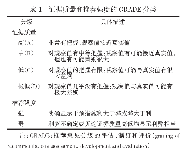
This consensus grades the quality of evidence and strength of recommendations for each clinical question using the Grading of Recommendations Assessment, Development and Evaluation (GRADE) method. The GRADE system classifies the quality of evidence into four levels: high (A), moderate (B), low (C), and very low (D), and the strength of recommendations into strong and weak (Table 1).
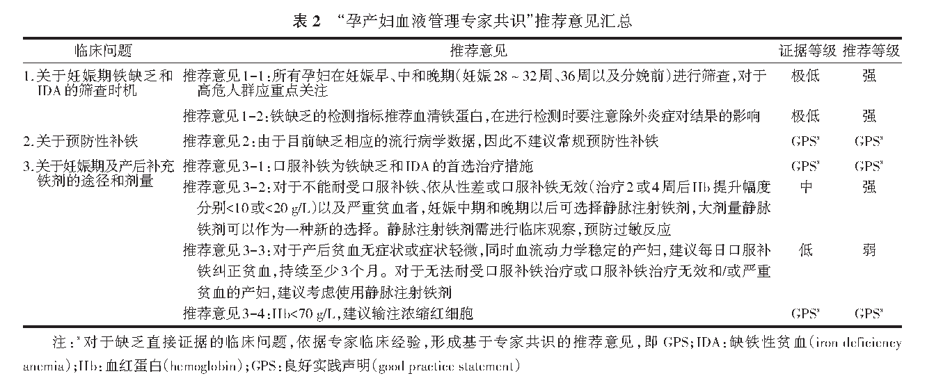
Summary of Recommendations in This Consensus
I. Strategies for Maternal Blood Management at Different Stages
1. Pregnancy
The core goal of patient blood management (PBM) in pregnancy is to increase maternal red blood cell reserves, early identify anemia and determine its etiology (e.g., iron deficiency anemia, thalassemia, vitamin B₁₂/folate deficiency, anemia of chronic disease), and correct it in a timely manner. Screening for anemia, standardized treatment, and follow-up are recommended during pregnancy and even the preconception period, with a focus on high-risk groups, including those on long-term vegetarian diets, underweight before pregnancy, chronic kidney disease, gastric ulcer, multiple pregnancies, and pregnant women complicated with obstetric bleeding disorders such as placenta accreta spectrum and placenta previa. Timely assessment and intervention reduce the incidence and severity of anemia and postpartum hemorrhage, and decrease the need for peripartum blood transfusion. Hemoglobin (Hb) levels should be rechecked before delivery, especially in pregnant women with anemia in pregnancy, to dynamically monitor anemia and enable timely correction.
2. Peripartum Period
The primary objectives of PBM in the peripartum period are to assess bleeding risk factors, increase maternal blood reserves, and implement measures to minimize peripartum blood loss, thereby reducing transfusion rates. Standardized obstetric delivery practices should be followed to reduce iatrogenic blood loss, active management of the third stage of labor should be performed to prevent postpartum hemorrhage, and blood loss should be accurately assessed. For women at high risk of obstetric bleeding, such as those with multiple pregnancies, placenta accreta spectrum, or placenta previa, thorough pre-delivery assessment and contingency planning are required. In institutions with available resources, intraoperative autologous blood transfusion may be prepared for cesarean delivery patients at risk of massive hemorrhage according to clinical circumstances. Individualized measures to reduce blood loss should be actively adopted for both vaginal and cesarean delivery, and a multidisciplinary team should be convened for collaborative management when necessary.
3. Puerperium
PBM in the puerperium aims to further reduce the risk of postpartum hemorrhage, improve maternal tolerance to anemia, restore red blood cell reserves to normal levels as soon as possible, and thereby reduce transfusion risks. Hemoglobin (Hb) levels should be measured promptly after delivery to assess the severity of anemia, followed by active correction and follow-up.
II. Management of IDA in Pregnancy
Anemia is a common complication of pregnancy and one of the core components of obstetric patient blood management (PBM). Iron deficiency is the leading cause of anemia in pregnancy, closely related to increased maternal and fetal iron requirements and physiological changes during pregnancy. Domestic research data show that the incidence of anemia in pregnancy is 17.3% in China, with an iron deficiency incidence as high as 45.6%. Existing evidence confirms that anemia and iron deficiency in pregnancy can have adverse short- and long-term effects on maternal and fetal health. In pregnant women, this can manifest as decreased exercise tolerance, fatigue, cognitive impairment, palpitations, headache, pallor, and reduced immunity, while increasing the risk of postpartum hemorrhage. Studies have shown that a 10 g/L decrease in prenatal Hb is associated with a 25% increase in the risk of postpartum hemorrhage, and severe anemia can lead to maternal shock and even death. For fetuses and neonates, moderate to severe anemia in pregnancy can affect their growth and development and is associated with increased risks of preterm birth, neonatal infectious diseases, and neurodevelopmental impairment.
Clinical Question 1: Timing of Screening for Iron Deficiency and IDA in Pregnancy
Recommendation 1-1: All pregnant women should be screened in the first, second, and third trimesters (28–32 weeks, 36 weeks of gestation, and before delivery), with special attention to high-risk groups. (Quality of evidence: very low; Grade of recommendation: strong)
Recommendation 1-2: Serum ferritin (SF) is recommended as the test for iron deficiency, and the influence of inflammation on results should be considered when performing the test. (Quality of evidence: very low; Grade of recommendation: strong)
Studies have shown that Hb concentrations start to decrease in the first trimester, reach a nadir in the second trimester, and gradually rise in the third trimester in healthy pregnant women without preconception iron deficiency, adapting to increased maternal blood volume and fetal iron requirements. Total iron requirements during pregnancy are approximately 1000 mg, increasing progressively with gestational age from 0.8 mg/day in the first trimester to 6.3 mg/day in the second and third trimesters. Therefore, pregnant women are a high-risk group for iron deficiency and iron deficiency anemia (IDA), making screening for iron deficiency and IDA clinically significant. However, there is currently a lack of research evidence on the optimal timing of screening for iron deficiency and IDA in pregnancy and its impact on maternal health and birth outcomes.
The definition of anemia in pregnancy varies slightly across countries. The WHO and the United States define anemia in pregnancy as Hb < 110 g/L in the first trimester, < 105 g/L in the second trimester, and < 110 g/L in the third trimester; the UK diagnostic criteria are Hb < 110 g/L in the first trimester and < 105 g/L in the second and third trimesters, based on Hb distribution levels in healthy pregnant women in different regions. China currently lacks large-scale local data and still uses Hb < 110 g/L as the unified diagnostic cutoff for anemia in pregnancy. It is also recommended that institutions with available resources measure serum ferritin (SF), with SF < 20 µg/L used to diagnose IDA. Pregnant women with risk factors for obstetric bleeding, such as long-term vegetarianism, underweight, chronic kidney disease, gastric ulcer, multiple pregnancies, and complications such as placenta accreta spectrum and placenta previa, should undergo targeted screening and management. The diagnostic threshold for SF in iron deficiency during pregnancy is not yet unified: the WHO recommends SF < 15 µg/L in the first trimester to indicate iron deficiency, the US, UK, and Australia use SF < 30 µg/L, and China recommends SF < 20 µg/L. All these criteria lack high-quality evidence-based support, and further research is needed to unify the diagnostic threshold for iron deficiency to promote the standardized management of iron deficiency and IDA in pregnancy.
The main examination for anemia in pregnancy is a complete blood count, and tests for iron deficiency mainly include bone marrow iron staining, serum ferritin (SF), total iron-binding capacity, and transferrin. SF is currently the most specific and minimally invasive biochemical indicator reflecting body iron stores, recommended by the WHO for assessing iron reserves, but interference from inflammatory conditions should be considered during testing.
Due to the lack of direct evidence, guidelines have not yet reached a consensus on the optimal timing of screening for IDA in pregnancy. It is recommended that all pregnant women undergo routine complete blood count testing to identify anemia early and determine if it is IDA via SF testing for timely intervention. Recommendations on screening timing vary across countries and regions, mainly based on physiological changes in pregnancy, iron requirements at each stage, and local prenatal care guidelines. The US recommends two Hb screenings: in the first trimester and 24–28 weeks of gestation; the UK and FIGO recommend screening at the first prenatal visit and 28 weeks of gestation; and the Asian Expert Consensus on the Management of Iron Deficiency Anemia in Obstetrics and Gynecology recommends screening at the first prenatal visit and early third trimester.
Combining the practical feasibility of anemia screening in pregnancy in China, aiming to minimize missed diagnoses, and referencing China’s current prenatal and postnatal care guidelines, this consensus recommends that all pregnant women be screened for anemia in the first, second, and third trimesters (28–32 weeks, 36 weeks of gestation, and before delivery). At present, there is controversy over whether routine iron deficiency screening is performed in pregnancy, with only some countries recommending routine SF testing in the first trimester. Given the uneven distribution of medical and health resources and medical cost burdens in China, some primary care institutions find it difficult to conduct routine SF testing, so this consensus only recommends it in institutions with available resources. It should also be noted that in areas with a high prevalence of thalassemia, thalassemia should be excluded during screening.
Clinical Question 2: Prophylactic Iron Supplementation in Pregnancy
Recommendation 2: Routine prophylactic iron supplementation is not recommended due to the lack of corresponding epidemiological data. (GPS)
There is no unified conclusion on whether routine prophylactic iron supplementation is needed for preconception and pregnant women. A 2023 meta-analysis including 8 randomized controlled trials (RCTs) with 2282 pregnant women without anemia or iron deficiency showed that daily oral iron supplementation during pregnancy reduced the risk of IDA at term (RR=0.51, 95% CI: 0.38–0.70; moderate certainty evidence) and low birth weight (RR=0.30, 95% CI: 0.13–0.68; moderate certainty evidence). A 2024 systematic review found that prophylactic iron supplementation reduced the incidence of anemia and iron deficiency at term, with a 70% decrease in term anemia (RR=0.30, 95% CI: 0.19–0.46), 67% decrease in term IDA (RR=0.33, 95% CI: 0.16–0.69), and 57% decrease in term iron deficiency (RR=0.43, 95% CI: 0.27–0.66), but the overall quality of included evidence was rated as low or very low.
In addition, some studies suggest that prophylactic iron supplementation may cause iron overload, leading to increased oxidative stress, adverse effects on pregnant women and offspring, and an increased risk of preterm birth, gestational diabetes mellitus, and preeclampsia. Studies have shown that serum ferritin (SF), serum iron, and transferrin saturation are significantly higher in women with gestational diabetes at 28–31 weeks of gestation compared with controls. Other studies have found that SF and serum iron concentrations are higher in patients with preeclampsia (median: 36.5 vs. 20.9 μg/L, P=0.019; 103.9 vs. 90.8 μg/dL, P=0.345), and SF > 40 μg/L is associated with preeclampsia (r=0.281, P=0.032).
Recommendations on prophylactic iron supplementation in pregnancy vary across countries. The WHO recommends 60 mg of elemental iron daily for pregnant women in areas with a high prevalence of anemia (≥40%); the US recommends low-dose iron supplementation from the first trimester to prevent anemia; the UK recommends 40–80 mg of elemental iron daily for pregnant women at risk of anemia; and FIGO recommends 30–60 mg of elemental iron daily for all pregnant women. Due to the lack of local research evidence in China, routine prophylactic iron supplementation is not yet implemented, and clinical decisions are still mainly based on Hb and SF results. During pregnancy, a balanced diet should be emphasized with diverse food intake and adequate minerals and vitamins; iron-rich foods such as animal blood, liver, and red meat should be increased, fruits and vegetables rich in vitamin C should be consumed to promote iron absorption, and co-administration with coffee and tea should be avoided to reduce inhibition of iron absorption.
Clinical Question 3: Routes and Doses of Iron Supplementation During and After Pregnancy
Recommendation 3-1: Oral iron supplementation is the first-line treatment for iron deficiency and IDA. (GPS)
Recommendation 3-2: For those who cannot tolerate oral iron supplementation, have poor adherence, or show no response to oral iron supplementation (Hb increase <10 g/L after 2 weeks or <20 g/L after 4 weeks of treatment), as well as those with severe anemia, intravenous iron may be considered after the second and third trimesters of pregnancy, and high-dose intravenous iron may be a new option. Clinical monitoring is required for intravenous iron administration to prevent allergic reactions. (Quality of evidence: moderate; Strength of recommendation: strong)
Recommendation 3-3: For postpartum women with asymptomatic or mild anemia and stable hemodynamics, daily oral iron supplementation is recommended for at least 3 months to correct anemia. For those who cannot tolerate oral iron supplementation, have poor adherence, or show no response to oral iron supplementation and/or have severe anemia, intravenous iron is recommended. (Quality of evidence: low; Strength of recommendation: weak)
Recommendation 3-4: For Hb <70 g/L, transfusion of packed red blood cells is recommended. (GPS)
Iron supplementation for pregnant women with IDA and iron deficiency in pregnancy mainly includes oral and intravenous iron supplementation. Given its good safety and cost-effectiveness, oral iron supplementation is recommended as first-line therapy by relevant guidelines and expert consensuses at home and abroad; intravenous iron may be used for pregnant women who cannot tolerate oral iron, have poor adherence, or show poor treatment response, or have severe anemia. In addition, the first trimester is a critical period for fetal organ differentiation and development, and there is insufficient safety data on intravenous iron at this stage, so oral iron is preferred in the first 3 months of pregnancy.
There is no unified consensus on iron supplementation doses for IDA in pregnancy, with recommended doses varying across guidelines and clinical practice. The UK guidelines recommend 40–80 mg of elemental iron daily, with Hb rechecked after 2–3 weeks to assess efficacy; for those who cannot tolerate or respond to oral iron, intravenous iron may be considered from the second trimester. The Asian Expert Consensus on the Management of Iron Deficiency Anemia/Iron Deficiency in Women proposes that daily oral elemental iron >60 mg may impair iron absorption in pregnant women, so alternate-day supplementation is recommended; for patients with IDA in the second and third trimesters with Hb <100 g/L after 2–3 weeks of oral iron or poor adherence, intravenous iron may be considered. For those diagnosed with IDA after 34 weeks of gestation and Hb <100 g/L, intravenous iron is recommended as first-line therapy to reduce transfusion risk. Chinese guidelines recommend 100–200 mg of elemental iron daily for IDA pregnant women, with Hb rechecked after 2 weeks to assess efficacy; for non-anemic pregnant women with SF <30 µg/L, 60 mg of elemental iron daily is recommended, with efficacy assessed after 8 weeks. For those who cannot tolerate, have poor adherence to, or show no response to oral iron, intravenous iron may be used after the second trimester. Transfusion of packed red blood cells is recommended for Hb <70 g/L; for Hb 70–100 g/L, transfusion decisions should be made based on factors such as surgery and cardiac function; iron supplementation (oral or intravenous) may be combined with transfusion.
A recent meta-analysis showed that compared with oral iron, intravenous iron achieved target Hb levels faster in pregnant women (OR=2.66, 95% CI: 1.71–4.15), with higher Hb levels after 4 weeks of treatment [WMD=0.84 g/dl (1 g/dl=10 g/L), 95% CI: 0.59–1.09], and lower incidence of adverse reactions (OR=0.35, 95% CI: 0.18–0.67). Other meta-analyses have also confirmed that intravenous iron has a faster onset and fewer adverse reactions, but the risk of allergic reactions remains. Studies on postpartum anemia show that compared with oral iron, intravenous iron significantly improves postpartum fatigue (SMD=−0.40, 95% CI: −0.62 to −0.18) and has a better effect on Hb elevation (MD=0.54 g/dl, 95% CI: 0.47–0.61), but the overall quality of evidence is moderate or low.
With the emergence of new intravenous iron preparations, iron is more tightly bound to carbohydrates, allowing controlled release and administration of high doses in a short time. Studies have shown that 1000 mg of ferric carboxymaltose in the second and third trimesters maintains iron reserves more stably and reduces repeated infusions; compared with oral iron, intravenous infusion of 1000 mg of ferric carboxymaltose over 15 minutes increases Hb levels faster and reduces the risk of adverse events. An RCT including 201 pregnant women with persistent iron deficiency at 14–21 weeks of gestation showed that a single infusion of 1000 mg of iron isomaltoside significantly increased Hb levels and improved fatigue symptoms compared with daily oral iron, with similar overall adverse reaction rates in both groups. Current studies have recommended intravenous iron as first-line therapy for iron deficiency in pregnancy, but pregnant women should be monitored during and within 30 minutes after intravenous iron administration to reduce the risk of allergic reactions. RCTs on postpartum anemia show that a single infusion of iron isomaltoside reduces physical fatigue scores at 12 weeks postpartum, corrects anemia better, and improves iron reserves without affecting milk iron content; both intravenous ferric carboxymaltose and iron isomaltoside are superior to oral iron in elevating Hb and improving iron metabolism, but some studies suggest that ferric carboxymaltose may increase the risk of hypophosphatemia, so the US guidelines recommend monitoring serum phosphate levels in patients treated with ferric carboxymaltose who are at risk of hypophosphatemia or require repeated dosing. China currently lacks research data on high-dose intravenous iron, and considering that oral iron is low-cost, convenient to use, safe, and effective, oral iron is still recommended as first-line therapy for iron deficiency and IDA in pregnancy; for those who cannot tolerate, have poor adherence to, or show no response to oral iron, intravenous iron may be used in the second and third trimesters.
After Hb returns to normal with IDA treatment, continued oral iron supplementation for ≥3 months is recommended to replenish body iron stores. It should be noted that excessively high Hb during pregnancy also increases the risk of pregnancy complications/adverse neonatal outcomes. A 2023 multicenter prospective study showed that compared with pregnant women with Hb 110 g/L in pregnancy, those with Hb 160 g/L had an increased risk of gestational hypertension (RR=1.67, 95% CI: 1.04–2.67), and those with Hb 170 g/L had more than a 2-fold increased risk (RR=2.29, 95% CI: 1.19–4.39); the risk of preterm birth and neonatal acute respiratory distress syndrome has a "U-shaped" relationship with gestational Hb levels, with significantly increased risks at Hb <100 g/L or Hb >145 g/L. Therefore, routine Hb monitoring is recommended during IDA treatment in pregnancy to avoid excessively high or low levels.
For women with postpartum hemorrhage or uncorrected anemia before delivery, Hb should be measured within 48 hours after delivery to assess anemia. For women with Hb <100 g/L and no obvious symptoms, daily supplementation of 100–200 mg of elemental iron is recommended, with Hb and SF rechecked after 3 months; for those who cannot tolerate or have no response to previous oral iron, intravenous iron is recommended to correct anemia.
Conclusion
In summary, obstetric patient blood management (PBM) is a patient-centered clinical practice throughout pregnancy, the peripartum period, and the puerperium, aiming to improve maternal red blood cell levels, identify and correct anemia in a timely manner, reduce blood loss and transfusion needs, and provide optimal treatment for pregnant women to improve pregnancy outcomes. The standardized management of anemia (mainly IDA) during pregnancy and the puerperium is an important component of obstetric PBM, with distinct management goals and focuses in each stage. It is expected that this consensus will provide a reference for maternal blood management in China and ultimately align with the goals of the World Health Organization (WHO) action framework.
Sources of Images and Text:中华预防医学会生命早期发育与疾病防控专业委员会, 中华医学会围产医学分会, 中华医学会妇产科学分会产科学组. 孕产妇血液管理专家共识[J]. 中华围产医学杂志, 2026, 29(2): 97-104. DOI: 10.3760/cma.j.cn113903-20250120-00033
Editor: Lily