
Chinese Voices | Team of Academician Huang Hefeng
Chinese Voices | Academician Huang Hefeng’s Team Publishes Landmark Findings in Nature: First Global Spatiotemporal Transcriptomic Atlas of Early Human Organogenesis
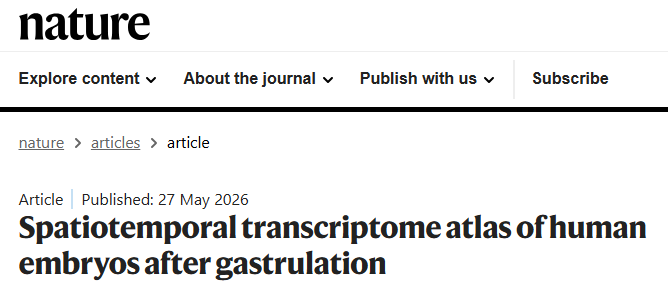
Recently, the team led by Academician Huang Hefeng of Zhejiang University published landmark findings in Nature. The team successfully constructed the world's first spatiotemporal dynamic transcriptomic atlas of human embryos during the post-gastrulation and early organogenesis stages. This breakthrough resolves a long-standing research challenge in the field, showcasing the cutting-edge scientific capabilities of China's obstetrics and gynecology community to the international academic world, while providing milestone data support for clinical and basic research in birth defect prevention, reproductive medicine, and fetal medicine.
Source: Nature
Research Background: Unlocking the "Black Box" of Embryonic Development
Weeks 4 to 8 post-fertilization (Carnegie Stages 12–23) represent the critical phase of human organogenesis, during which core tissues and organs—such as the heart, brain, liver, and kidneys—gradually differentiate and develop. Abnormal embryonic development during this period is the primary trigger for various birth defects, including congenital heart disease and neurodevelopmental disorders.
Due to multiple constraints such as sample accessibility and technical limitations, the gene expression, cell differentiation, and molecular regulatory mechanisms during this stage have remained systematically unresolved, creating a persistent blind spot in developmental biology and obstetrics. Clarifying the developmental laws of this stage holds irreplaceable clinical value for interpreting the etiology of birth defects, guiding early pregnancy care, and advancing fetal disease diagnosis and treatment.
Research Strategy: Multi-Technology Integration for Systematic Analysis
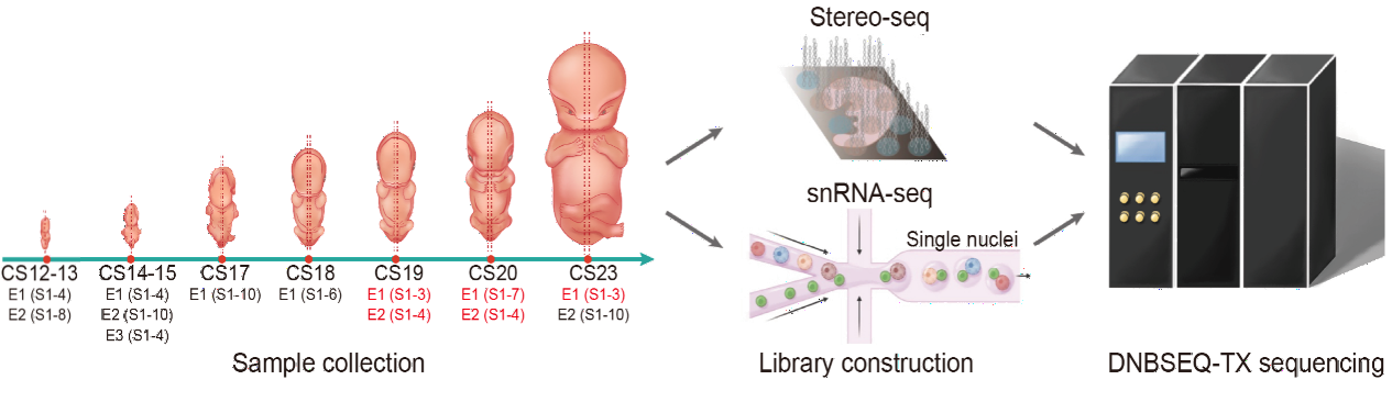
Spanning five years and strictly adhering to medical ethics standards, the research team innovatively combined Stereo-seq high-resolution spatial transcriptomics with single-nucleus RNA sequencing, overcoming the shortcomings of traditional detection methods.
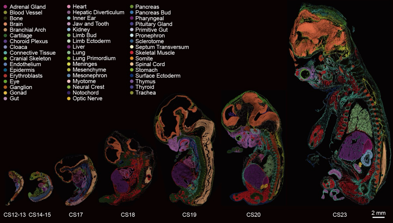
The study analyzed 13 human embryos corresponding to the target developmental stages, conducting analysis on 77 sagittal sections. The team finely delineated 50 organ anatomical regions and 198 molecular substructures, fully reconstructing the dynamic characteristics of cell migration, differentiation, and gene expression during embryogenesis. Additionally, the team constructed the first organ-level atlas of allele-specific imbalanced expression in human embryos, validating known imprinted genes and screening a batch of novel candidate imprinted genes, thereby further refining the framework of embryonic epigenetics research.
Source: Nature
Figure Note: Schematic diagram of the research workflow, covering the entire process from sample collection and library construction to sequencing analysis.
Core Breakthroughs: New Discoveries Reshaping International Understanding
Identifying Key Genes for Cardiac Development, Advancing Congenital Arrhythmia Research
The study pinpointed two previously uncharacterized genes, RORAand KIAA1324L, confirming their roles as core regulatory genes for the differentiation of sinoatrial node cells (the heart's "natural pacemaker") and the maintenance of heart rate homeostasis. These conclusions were validated through zebrafish and mouse experiments, opening new avenues for exploring the pathogenesis of congenital arrhythmias and developing targeted therapies.
Source: Nature
Figure Note: Spatiotemporal atlas of human embryo organogenesis at CS12-23. Different colors mark the distribution characteristics of 50 organs and 198 molecular substructures.
Revising Neuronal Developmental Timelines and Analyzing Neurological Disease Mechanisms
This study updates traditional textbook views: inhibitory neuron markers were detected as early as 4–5 weeks post-fertilization (CS12–13), and excitatory neuron markers appeared by 7 weeks (CS19)—both significantly earlier than previous conclusions.
Furthermore, the study identified a HMGA2-centric gene regulatory network highly associated with genes linked to intellectual disability, providing crucial clues for research into neurodevelopmental disorders such as autism, epilepsy, and fetal intellectual developmental deficits.
Elucidating the Window of Susceptibility to Maternal Infections, Guiding Early Pregnancy Protection
The team systematically analyzed the distribution of receptors for common pathogens—including cytomegalovirus, Zika virus, hepatitis B virus, and SARS-CoV-2—within embryonic tissues. They confirmed that these viral receptors exhibit both organ-specificity and developmental stage-specificity. This work explains the "window of susceptibility effect" at the molecular level, clarifying why infections at different gestational weeks or from different viruses specifically damage certain fetal organs, thus providing a scientific basis for clinical risk assessment and maternal-fetal protection.
Highlighting Limitations of Model Organisms, Standardizing Basic Research and Translation
By comparing gene expression data between human and mouse embryos, the study found significant discrepancies in the timing of disease-associated gene expression. This result indicates that experimental conclusions drawn from model organisms like mice cannot be directly extrapolated to humans, establishing a critical reference standard for basic reproductive research, drug development, and clinical translation.
Research Value: Scientific Breakthrough and Clinical Significance
This achievement has been evaluated by international peers as a milestone in developmental biology. From a basic research perspective, the atlas completely fills a gap in our understanding of early human organogenesis, upgrading the field's knowledge from fragmented observation to a holistic, dynamic, and refined molecular panorama. From a clinical perspective, the findings provide molecular support for first-trimester prenatal screening and risk assessment of fetal developmental anomalies, facilitating the shift of birth defect prevention upstream.
Future Outlook: Deepening the Translation from Bench to Bedside
This study stands as a milestone in human embryonic development, with multiple original discoveries laying a solid theoretical foundation for birth defect prevention, fetal medicine, and reproductive biology. Moving forward, Academician Huang Hefeng’s team will continue to integrate scientific research with clinical practice, further exploring embryonic regulatory mechanisms and accelerating the translation of research outcomes to achieve early warning and precise intervention for fetal developmental abnormalities. This work also fully demonstrates the research strength of China's obstetrics and gynecology community, contributing vital "Chinese wisdom" to global scientific endeavors.
Source: Pan, J., Li, Y., Lin, Z. et al. Spatiotemporal transcriptome atlas of human embryos after gastrulation. Nature(2026). https://doi.org/10.1038/s41586-026-10545-0
[Disclaimer: This study is basic scientific research based on human embryonic samples and model animal experiments. The clinical translation of related conclusions requires further validation. This content is for academic exchange and popular science reference only and does not constitute any clinical diagnostic or treatment advice.]
Editor: Lily