





















Expert Consensus on Emergency Management of Critical Illness in Pregnancy and Childbirth (2026 Edition): 12 Key Recommendations
China has achieved remarkable progress in maternal critical care, with the maternal mortality ratio declining sharply from 53 per 100,000 in 2000 to 14.3 per 100,000 in 2024. However, with the optimization of fertility policies, the incidence of pregnancy complications and comorbidities has risen, leading to a surge in obstetric critical care cases and posing severe challenges to maternal-fetal safety. While the 2024 edition of the Chinese Expert Consensus on Early Identification of Obstetric Critical Illness provided a foundation, several key clinical challenges remained unresolved in practice:
1. Lack of clear standardized pathways for emergency management following early identification of obstetric critical illness.
2. Uncertainty regarding the rational integration of real-time vital sign monitoring with auxiliary diagnostics to dynamically optimize resuscitation protocols.
3. Difficulty in implementing efficient interventions within narrow therapeutic windows for hemodynamically unstable patients to reduce adverse outcomes.
To address these gaps, a multidisciplinary expert panel—comprising specialists in obstetrics & gynecology, critical care, laboratory medicine, ultrasound, and radiology—was convened to develop this consensus. Drawing on the latest global evidence and local clinical realities, the panel formulated the Expert Consensus on Emergency Management of Critical Illness in Pregnancy and Childbirth (2026 Edition).
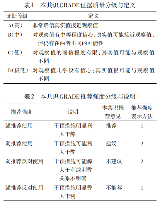
The quality of evidence and strength of recommendations herein are graded according to the Grading of Recommendations Assessment, Development, and Evaluation (GRADE) system (Tables 1 and 2).
Identification and Emergency Management of Critically Ill Pregnant Women
A Maternal Near Miss (MNM) is defined as a pregnant or postpartum woman (within 42 days of delivery) who experiences a life-threatening pregnancy-related complication or pre-existing condition but survives following intensive medical intervention.
Recommendation 1:
Utilize the Airway, Breathing, Circulation, Disability, Exposure (ABCDE) algorithm for rapid assessment to identify immediately life-threatening conditions. Simultaneously initiate emergency interventions, activate emergency calls, and mobilize multidisciplinary team (MDT) collaboration. (Evidence Level: A; Recommendation Strength: 1)
01 Rapid Assessment
Upon suspicion of critical illness, initiate the ABCDE assessment immediately while summoning the emergency team.
• Airway: Assess spontaneous ventilation and ability to speak. In unconscious patients, inspect the oropharynx for obstruction (e.g., due to eclampsia, aspiration, or anaphylaxis).
• Breathing: Observe for cyanosis or accessory muscle use. Respiratory rates <6 or >40 breaths/min, or signs of respiratory distress/failure, necessitate immediate intervention. Common obstetric causes include pneumonia, pulmonary edema, pulmonary embolism (PE), and amniotic fluid embolism (AFE). Indications for intubation and mechanical ventilation include severe respiratory failure, persistent hypoxemia, respiratory acidosis, or altered mental status.
• Circulation: Monitor for hypotension, arrhythmias, cold clammy skin, delayed capillary refill, weak pulses, and oliguria. Common triggers include hemorrhage, sepsis, PE, heart failure, and AFE. If cardiac arrest is suspected (unresponsiveness, no breathing/apnea, no pulse), initiate high-quality Cardiopulmonary Resuscitation (CPR) immediately. For pregnancies ≥20 weeks or uterine fundus at/above the umbilicus, perform manual left uterine displacement to relieve aortocaval compression. If return of spontaneous circulation (ROSC) is not achieved after 4 minutes of CPR, proceed to perimortem cesarean delivery.
• Disability: Assess consciousness (AVPU scale) and pupillary response. Altered mental status may indicate metabolic disturbances (hypo/hyperglycemia, electrolyte imbalance, organ failure). Perform point-of-care testing (POCT) including finger-stick glucose and arterial blood gas (ABG) analysis.
• Exposure: Conduct a brief history and physical exam to identify bleeding, concealed hemorrhage, or infection sources while preventing hypothermia.
Recommendation 2:
Initial management prioritizes stabilizing vital signs while concurrently identifying the underlying etiology. Immediately secure the airway, administer oxygen, establish venous access, and monitor dynamically to adjust the treatment plan. (Evidence Level: A; Recommendation Strength: 1)
02 Initial Emergency Interventions
The primary goal is to monitor the "Eight Vital Signs" (consciousness, respiration, pulse, blood pressure, pupils, skin/mucous membranes, temperature, urine output) and adhere to the principle of "stabilize first, diagnose second."
1. Continuous Monitoring: Monitor vital signs nearly continuously during hemodynamic instability; once stable, monitor at intervals no longer than 5 minutes.
2. Oxygenation and Ventilation: Maintain SpO₂ > 95% and PaO₂ > 60 mmHg. Administer supplemental oxygen via face mask (5–10 L/min). If intubation is impossible, use a bag-valve-mask (BVM) with a 1:1.5–2 inspiratory-to-expiratory ratio, tidal volume of 400–600 mL, and rate of 10–12 breaths/min. Synchronize assistance with spontaneous breaths. Involve Anesthesiology and ICU early.
3. Intravenous Access: Establish large-bore (16–18G) peripheral IV access in the upper extremities. If unsuccessful, insert two smaller catheters or use ultrasound guidance. Initiate crystalloid infusion (balanced salt solution). Consider central venous or intraosseous access if peripheral access fails. Falling heart rate, rising blood pressure, and increasing urine output indicate successful resuscitation.
4. Temperature Management: Maintain core temperature between 36.5–37.5°C. Use forced-air warming blankets and fluid warmers. Transfuse blood products via fluid warmers. Manage hyperthermia promptly to prevent vasoconstriction and microcirculatory compromise.
Dynamic Assessment and Optimization
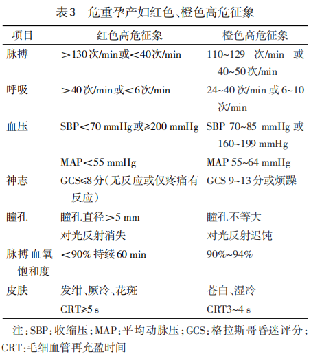
Pregnancy induces physiological changes (hypervolemia, hypercoagulability) altering normal lab values. Refer to WHO criteria to identify "Red" and "Orange" alert signs (Table 3) immediately.
Recommendation 3:
Rapidly identify high-risk warning signs. Strictly enforce the first-responsibility system. Report cases immediately to the institutional Maternal Critical Care Team and the Regional Maternal Critical Care Center. Follow standardized protocols for referral and transfer. (Evidence Level: B; Recommendation Strength: 2)
Recommendation 4:
Conduct individualized comprehensive assessments post-resuscitation, including history, physical exam, and targeted diagnostics (ECG, POCUS, labs). Address critical lab values promptly and adjust the management plan dynamically. (Evidence Level: C; Recommendation Strength: 2)
01 Initial Comprehensive Assessment
1. History: Obtain focused history from patient/surrogates regarding prodromal symptoms, comorbidities, and prior tests. Prioritize ruling out lethal conditions: headache/vomiting (intracranial pathology, hypertensive emergency); chest/back pain (aortic dissection, ACS); abdominal pain (hepatic/rupture, uterine rupture).
2. Physical Exam: Assess posture, neck stiffness, mental status, pupils, skin, and edema. Perform cardiopulmonary and abdominal exams. Obstetric exam: fundal height, uterine tone, contractions, and fetal status.
3. Auxiliary Tests: Do not rely solely on lab results due to reporting delays; correlate with clinical trends.
◦ Immediate (0–15 min): Finger-stick glucose, ABG (Lactate, BE, PaO₂/FiO₂, electrolytes, Hgb), ECG.
◦ Urgent (15–60 min): CBC, Coagulation panel (PT, APTT, Fib), CMP (LFTs, RFTs, lytes), cardiac biomarkers (Troponin, CK-MB, BNP/NT-proBNP), sepsis markers (PCT, CRP), amylase/lipase (if pancreatitis suspected).
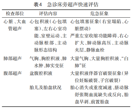
◦ Bedside Ultrasound (POCUS): Perform a sequential "Cardiac-Lung-Abdominal" scan (Table 4). Identify ectopic pregnancy, rupture, abruption, previa, and assess fetal status. If free fluid + hemodynamic instability, perform diagnostic paracentesis/culdocentesis. Decide on expedited delivery based on maternal/fetal status.
Recommendation 5:
Perform finger-stick glucose testing immediately. Glucose <3.3 mmol/L or >13.9 mmol/L constitutes a warning threshold requiring urgent intervention. Adjust monitoring frequency based on acuity and individualize glycemic targets. (Evidence Level: A; Recommendation Strength: 1)
4. Glucose Monitoring: Treat hypoglycemia (<3.3 mmol/L) with 50% dextrose IV push; monitor K⁺ closely. Treat hyperglycemia (>13.9 mmol/L) with insulin to target <10.0 mmol/L. Monitor hourly during instability, then q1–2h during stabilization, and q2–4h when stable.
Recommendation 6:
Obtain ABG analysis promptly to assess pH, Lactate, BE, PaO₂/FiO₂, electrolytes, and Hgb. Lactate ≥2.0 mmol/L is an independent predictor of poor prognosis. Address critical values immediately and adjust resuscitation accordingly. (Evidence Level: B; Recommendation Strength: 1)
5. Blood Gas Analysis:
◦ Acid-Base: pH <7.2 or >7.6 requires intervention. Calculate PaO₂/FiO₂ (RI <300 mmHg = RF; <200 mmHg = severe RF). BE ≤ -10 mmol/L indicates severe metabolic acidosis.
◦ Lactate: ≥2.0 mmol/L (hyperlactatemia); ≥4.0 mmol/L (critical value). A decrease of ≥10% over 2 hours suggests improved perfusion. Lactate >8.0 mmol/L carries a very high mortality risk. Correlate lactate with BE to differentiate types of shock.
◦ Electrolytes: K⁺ <2.5 or >6.0 mmol/L requires ECG correlation and urgent correction. Ionized Ca²⁺ <1.5 mmol/L impairs coagulation/contractility; replete. Mg²⁺: Therapeutic range 1.8–3.0 mmol/L; Toxicity >3.5 mmol/L. Monitor Na⁺ closely to avoid osmotic demyelination.
◦ Hemoglobin: Critical value <50 g/L. Target Hgb ≥70 g/L (low bleed risk) or ≥80 g/L (high bleed risk/active bleeding).
Recommendation 7:
Perform a comprehensive reassessment 1 hour after initiating resuscitation. Prioritize screening for the Lethal Triad (Hypothermia, Acidosis, Coagulopathy). Escalate care to MDT mode for critically ill patients. (Evidence Level: B; Recommendation Strength: 1)
02 One-Hour Dynamic Assessment
Reassess symptoms, vitals, and lab results (ABG, CBC, Coags) after 1 hour of treatment.
1. The Lethal Triad:
◦ Hypothermia: Core Temp <35°C.
◦ Acidosis: pH <7.2, Lactate >4.0 mmol/L.
◦ Coagulopathy: PT >18s (INR >1.5), APTT >60s, Fib <1.5 g/L, PLT <50×10⁹/L.
◦ Red Alert Criteria: Hgb drop >10 g/L/hr; RBC transfusion >4 units/hr (>10 units/24hr); Lactate drop <10% in 2hr; Urine <30 mL/hr x 2hr; PLT <50×10⁹/L; Fib <1.5 g/L + D-dimer >10–20 mg/L (or TEG LY30 >7.5%).
2. Severe Organ Dysfunction: Monitor for hepatic (TBil ≥100 μmol/L + AST/ALT >1000 U/L), renal (Cr ≥300 μmol/L), thyroid (Thyrotoxicosis/Myxedema Crisis), pancreatic (Amylase >1000 U/L), and cardiac (Trop >1.0 μg/L, NT-proBNP >1400 ng/L) dysfunction. Consult subspecialists immediately. Note: Lab results reflect past status; continuous clinical assessment is paramount.
Recommendation 8:
If severe hepatic, renal, or thyroid dysfunction is detected, initiate targeted interventions immediately and monitor organ function dynamically. (Evidence Level: B; Recommendation Strength: 1)
Recommendation 9:
If transport for off-bed imaging is necessary, weigh diagnostic benefit against transport risk rigorously. Select appropriate modalities and ensure continuous maternal life support during transit. (Evidence Level: B; Recommendation Strength: 1)
03 Imaging Studies
Prioritize life support over diagnostics. Avoid transporting unstable patients. If imaging is essential after initial stabilization, do not delay due to radiation concerns (inform patient/family, shield fetus). Ensure trained personnel and portable equipment accompany the patient.
• Indications for Emergency CT (<30 min from order to scan): Suspected PE (hypoxia/shock), intracranial hemorrhage (coma), stroke (neuro deficits), aortic dissection (tearing chest pain/hypotension).
• Modalities: Non-contrast CT (ICH/trauma); CTA (vascular occlusion/stroke); CTPA (PE); Whole-aorta CTA (dissection).
• MRI: Reserved for stable patients due to long duration and monitoring difficulties; useful for CNS/soft tissue characterization.
Classification, Early Recognition, and Management of Shock
Shock is a life-threatening circulatory failure. Major types include hypovolemic (primarily hemorrhagic), distributive (septic/anaphylactic), cardiogenic, and obstructive. Hemorrhagic and septic shock are most prevalent in obstetrics. Early recognition during the compensatory phase is critical to prevent progression to MODS.
Recommendation 10:
Recognize early signs of shock. Upon detecting hemodynamic instability (HR >100 bpm, SBP <100 mmHg, DBP <60 mmHg), activate the emergency protocol immediately. Monitor Capillary Refill Time (CRT) and hourly urine output continuously while stabilizing vitals and identifying the etiology. (Evidence Level: A; Recommendation Strength: 1)
01 Early Recognition and Diagnosis
Diagnosis combines clinical signs, hemodynamics, and biochemistry.
1. Hypotension: SBP <90 mmHg or MAP <70 mmHg. Note: Hypertensive patients may not appear hypotensive.
2. Hypoperfusion: Pale/cool skin, Urine <0.5 mL/kg/h, Altered mental status.
3. Hyperlactatemia: Indicates cellular hypoxia (may precede BP drop).
Intervention Goal: Restore perfusion and oxygenation; use lactate trends to guide therapy.
02 Management of Compensatory Phase and Hemodynamic Assessment
Principles: Stabilize hemodynamics; initiate fluid resuscitation pending diagnosis; treat underlying cause; use vasopressors (adrenergic agonists) if fluids fail.
• CRT: Normal 1–2s; >3s indicates hypoperfusion (early warning sign).
• Urine Output: <0.5 mL/kg/h or <30 mL/h indicates inadequate perfusion.
03 Emergency Management of Common Obstetric Shock
1. Hemorrhagic Shock
Postpartum hemorrhage (PPH) is the leading preventable cause of maternal death. Physiological hypervolemia masks early signs; significant blood loss (≈1000 mL) may occur before instability manifests. Loss >1500 mL leads to agitation, tachypnea, weak pulses, and cool clammy skin (compensatory phase). Without intervention, decompensated shock and MODS ensue rapidly.
• Early Identification (WHO 2025): Blood loss ≥300 mL + Hemodynamic instability (HR >100, SBP <100, DBP <60, SI >0.9). HR rise and SI >0.9 are sensitive early indicators.
• Goals: HR <100, SBP >100, Urine >30 mL/h, Hct >30%.
Recommendation 11:
Median time from hemorrhage onset to death is only 2 hours. Recognize compensated shock early. Implement standardized interventions within the "Golden Hour," prioritizing hemorrhage control, early tranexamic acid (TXA), and component blood transfusion to reverse the Lethal Triad. (Evidence Level: A; Recommendation Strength: 1)
• Golden Hour Protocol (Dynamic, not rigid):
◦ First 20 min: Etiology & Pharmacologic Control (4T's: Tone, Trauma, Tissue, Thrombin).
◦ Second 20 min: Mechanical Hemostasis (Balloon tamponade, sutures, vessel ligation, interventional radiology). Administer TXA within 3 hours of onset + Calcium.
◦ Third 20 min: Surgical Intervention/Damage Control. Hysterectomy if bleeding uncontrolled. In resource-limited settings, consider early hysterectomy.
• Decompensated Shock/MODS: Aggressive correction of coagulopathy. Consider Damage Control Surgery (packing, temporary closure) + Massive Transfusion Protocol (MTP) + MDT support.
2. Septic Shock
Current scoring systems (MEWS, qSOFA, SOFA, NEWS2) have limitations in pregnancy. Rely on clinical synthesis.
• Red Flag Criteria:
1. Neuro: New lethargy, confusion.
2. Circulatory: SBP ≤90 or drop >40 mmHg, mottling, CRT >3s, HR ≥130 bpm.
3. Respiratory: RR ≥25 bpm, SpO₂ <92% (room air).
4. Metabolic/Renal: Lactate >2 mmol/L, Oliguria (<0.5 mL/kg/h x 2hr).
5. Temperature: <35°C (high risk) or >39°C.
Recommendation 12:
Implement the "Golden Hour" bundle for septic shock: Measure lactate, obtain cultures, initiate broad-spectrum antibiotics, and commence fluid resuscitation to maintain MAP ≥65 mmHg. Use vasopressors (Norepinephrine first) if fluids fail. Simultaneously eradicate the source of infection. (Evidence Level: A; Recommendation Strength: 1)
• Golden Hour Bundle:
1. Assessment: Monitor vitals, ABG, CBC, Coags, LFTs/RFTs. Obtain cultures (blood x2, urine, swabs) BEFORE antibiotics.
2. Antibiotics: Administer IV within 1 hour of recognition.
3. Fluids: 30 mL/kg crystalloids (e.g., balanced salt). Consider bolus (10 mL/kg over 30 min) then titrate based on dynamic assessments (Passive Leg Raise, Fluid Challenge) to avoid overload.
4. Vasopressors: Norepinephrine to target MAP ≥65 mmHg if fluids fail.
• Source Control: Terminate pregnancy/drain abscess/remove infected devices urgently. Debride surgically once stable if needed. Consider mNGS for culture-negative sepsis.
Summary
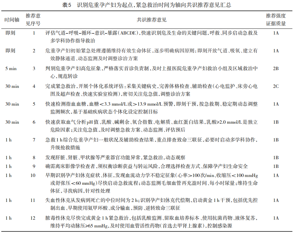
This consensus emphasizes early identification, a timeline-driven approach, and dynamic management to reduce maternal morbidity and mortality. It underscores the "Save the Mother First" principle, rigorous monitoring of the "Eight Vital Signs," prompt response to critical lab values, and MDT collaboration. Recognizing compensated shock and adhering to the "Golden Hour" paradigm are pivotal to reversing adverse outcomes. A post-event review (debriefing) is recommended to refine institutional protocols continually. (See Table 5 for a summary of all recommendations).
(Image 4)
Disclaimer: This content is derived from the Expert Consensus on Emergency Management of Critical Illness in Pregnancy and Childbirth (2026 Edition) and is intended for medical professional education only. It does not constitute clinical diagnosis, treatment advice, or medication guidance. Clinicians must formulate individualized treatment plans under the guidance of professional physicians based on specific patient conditions, institutional resources, and current clinical guidelines.
Source: Society of Obstetric and Perinatal Medicine, China International Exchange and Promotive Association for Medical and Health Care. Expert consensus on emergency management of critical illness in pregnancy and childbirth (2026 edition) [J]. Chinese Journal of Practical Gynecology and Obstetrics, 2026, 42(6): 619-627.
Editor: Huo Pan