





















Pelvic nerve injury is a common complication following radical hysterectomy, radical rectal cancer resection, and sacrococcygeal teratoma resection. It can directly induce neurogenic bladder, leading to loss of voluntary voiding, urinary retention, recurrent urinary tract infections (UTIs), and even progressive renal impairment, severely compromising quality of life and life safety.
Historically, intradural nerve transfer (the Xiao procedure) has been utilized for neurogenic bladder caused by spinal cord injury or spina bifida. However, as this approach involves the spinal canal, it cannot be applied to peripheral-type neurogenic bladder resulting from pelvic nerve transection during pelvic surgery.
The obturator nerve is a robust somatic nerve located anatomically adjacent to the pelvic nerves, providing a sound anatomical basis for transfer. Based on original clinical research data published in International Urology and Nephrology, this study systematically evaluates the efficacy of obturator-to-pelvic nerve transfer in restoring bladder sensation, motor function, and voluntary voiding via somatic-to-autonomic nerve regeneration.
Figure 1.
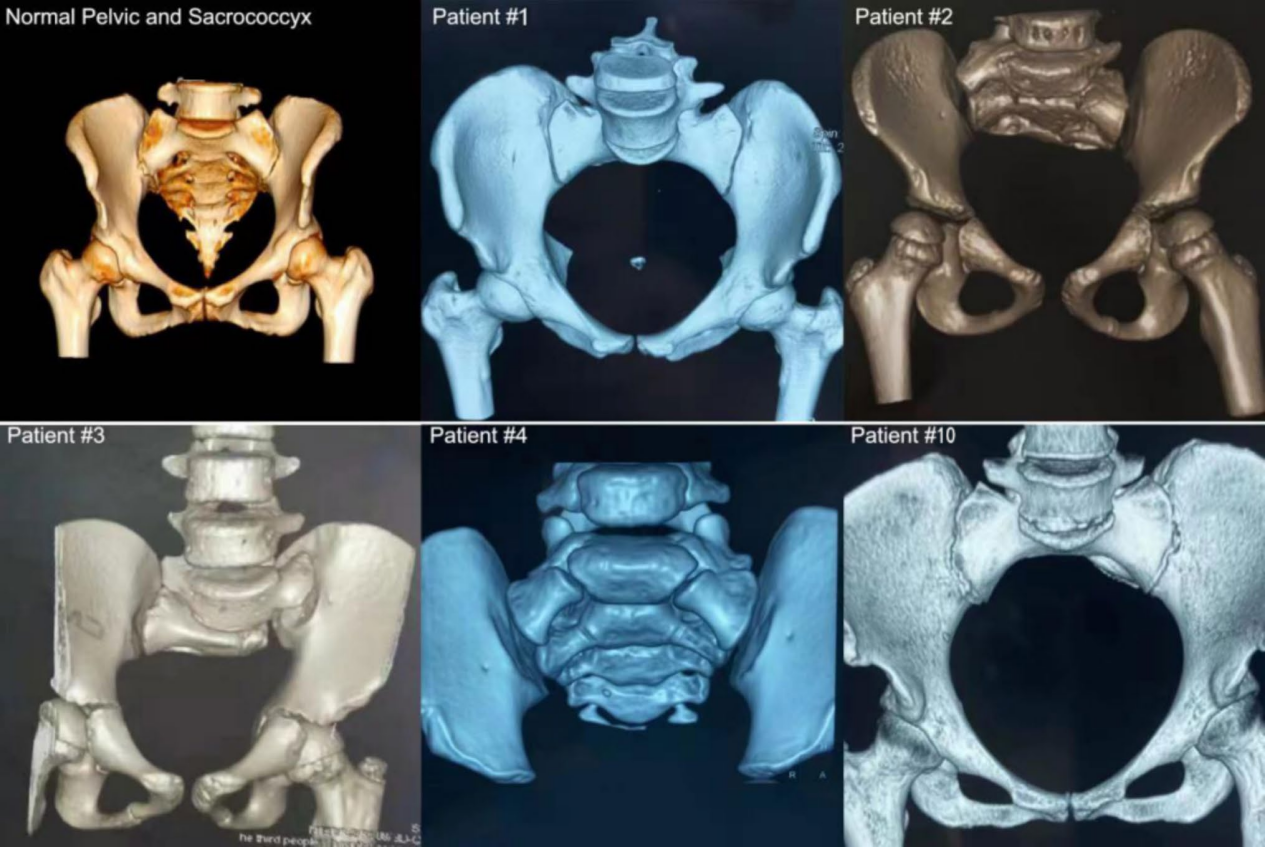
Comparison of pelvis in a normal individual versus a patient post-sacrococcygeal teratoma resection.
(Source: International Urology and Nephrology)
Materials and Methods
1. Study Population
This prospective interventional case series included 11 patients (2 males, 9 females), aged 4 to 58 years. Primary surgical pathologies included sacrococcygeal teratoma resection (n=5), presacral teratoma resection (n=2), and radical hysterectomy (n=4).
2. Surgical Technique
All procedures were performed under general anesthesia via a lower midline abdominal incision accessing the paravesical space. The obturator nerve was dissected, and the ventral one-third of its fascicles were harvested and coapted end-to-end with the distal stump of the transected pelvic nerve. For patients with severe concomitant constipation, a Y-shaped anastomosis was performed between the obturator nerve and both the vesical and rectal branches of the pelvic nerve.
Figure 2.
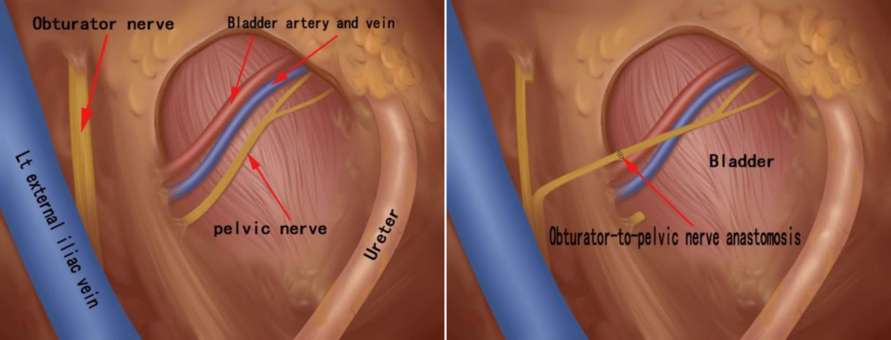
A. Schematic diagram of the anatomy of the left obturator nerve and pelvic nerve within the pelvic cavity.
B. Schematic diagram of obturator nerve-pelvic nerve transfer anastomosis.
Figure 3.
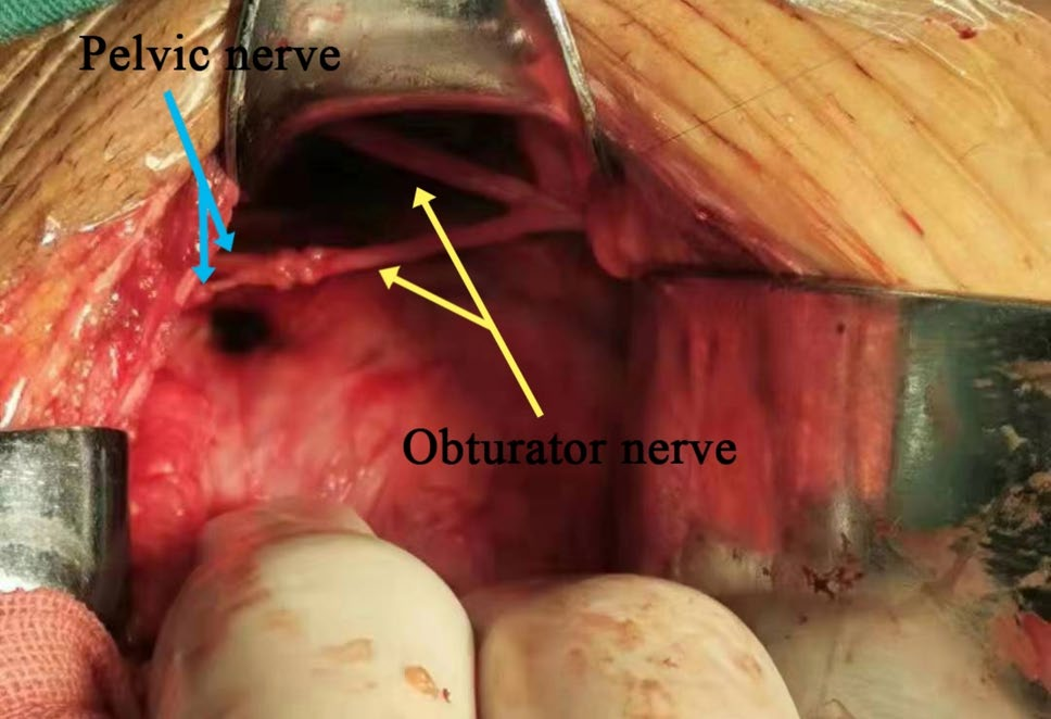
Intraoperative photograph showing the anastomosis between the right obturator nerve and the vesical and intestinal branches of the pelvic nerve.
3. Outcome Measures
Urodynamic studies were conducted preoperatively and at 18 months postoperatively. Parameters included voided volume, post-void residual (PVR) volume, detrusor systolic pressure, bladder compliance, and bladder sensation. UTI incidence and serum creatinine levels were recorded. Safety was assessed by monitoring surgical complications and lower limb function.
4. Statistical Analysis
Data were analyzed using paired t-tests. A P-value < 0.05 was considered statistically significant.
Results
1. Recovery of Voiding Function
At baseline, none of the 11 patients had voluntary voiding ability. At follow-up, 9 out of 11 patients (81.8%) regained spontaneous urination within 3–6 months post-surgery and no longer required catheterization. One patient showed no improvement, and one withdrew due to recurrence and metastasis of cervical cancer.
2. Urodynamic and Renal Improvement
The 9 patients who recovered voiding showed significant improvements:
Voided Volume: 0 ml → 285 ± 5.8 ml (P< 0.001)
Post-Void Residual (PVR): 240 ± 141.4 ml → 23 ± 18.6 ml (P< 0.01)
Detrusor Systolic Pressure: 3 ± 3.3 cmH₂O → 39 ± 6.5 cmH₂O (P< 0.001)
Bladder filling sensation returned, UTIs significantly decreased, and renal function improved markedly.
Figure 4.

Baseline characteristics of patients and changes in urination-related parameters between preoperative status and 18 months postoperative.
3. Urodynamic Cystometrogram (CMG) Patterns
Postoperatively, bladder compliance and capacity improved significantly, and the detrusor muscle regained effective contraction.
Figure 5.
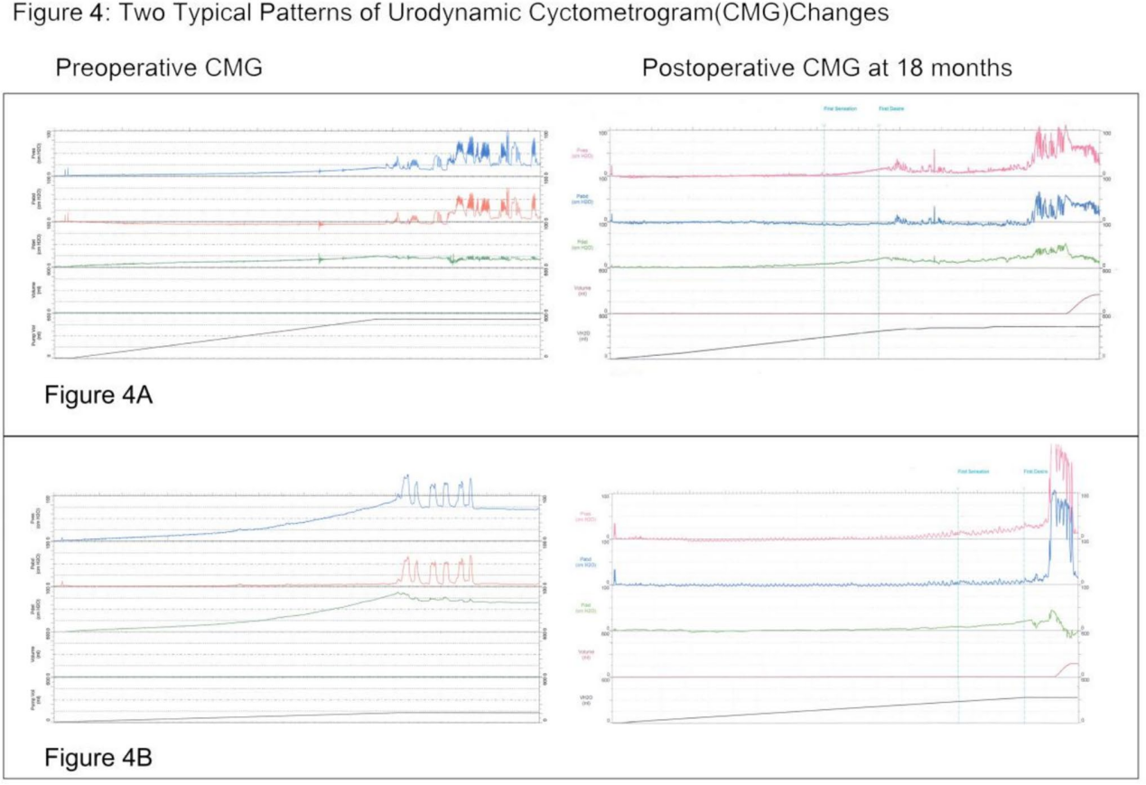
Typical changes in urodynamic cystometrogram (CMG) patterns before surgery and 18 months after obturator nerve-pelvic nerve transfer.
4. Safety
The procedure was technically feasible and safe. No perioperative complications or deficits in lower limb motor function were observed.
Discussion
1. Anatomy and Mechanism
Radical pelvic surgery often damages the pelvic nerves while preserving the pudendal nerve, primarily causing urinary retention. Obturator nerve transfer facilitates somatic-to-autonomic regeneration, reconstructing bladder innervation. Due to the short distance for nerve regeneration, functional recovery occurs rapidly within 3–6 months, significantly faster than intraspinal nerve transfer.
2. Distinction from the Traditional Xiao Procedure
The traditional Xiao procedure involves intradural lumbosacral nerve transfer within the spinal canal and is suitable for patients with spinal cord injury or spina bifida who often present with incontinence. In contrast, this technique is a peripheral nerve repair that does not involve the spinal canal, offering higher safety. It specifically targets peripheral-type neurogenic bladder caused by pelvic nerve transection, making the two procedures complementary in indication.
3. Clinical Value
This study provides the first human evidence that obturator nerve transfer effectively restores somato-autonomic innervation of the bladder, recovering both sensation and contraction. The procedure is straightforward and can be safely performed even in scarred pelvic tissues, making it suitable for early intervention post-pelvic surgery to prevent detrusor fibrosis.
4. International Validation
Recent clinical research from Massachusetts General Hospital (Harvard University) published in Neurosurgeryreported a successful case of bladder function restoration in a patient with sacrococcygeal tumor resection via obturator-to-pelvic nerve anastomosis. The technique and outcomes are consistent with the 5 cases of successful rescue reported by Professor Chuan-Guo Xiao in this study, further validating the international feasibility of this approach.
5. Precautions
Contraindications include bladder capacity >800 ml and tumor recurrence. Early nerve transfer is recommended to avoid detrusor fibrosis, which may compromise efficacy.
Conclusion
Obturator-to-pelvic nerve transfer is an effective surgical option for neurogenic bladder following radical hysterectomy or sacrococcygeal teratoma resection, achieving an 81.8% recovery rate of voluntary voiding. This technique significantly restores bladder sensation and contractility, reduces residual urine and UTIs, protects renal function, and may improve neurogenic bowel function. Supported by international validation, this procedure is safe, minimally invasive regarding limb function, and represents a preferred treatment for peripheral pelvic nerve injury-induced neurogenic bladder.
Source:
Xiao, X.Y., Hou, T., Li, B. et al. Obturator-to-pelvic nerve rerouting to restore bladder function after radical pelvic nerve injury. Int Urol Nephrol(2026). https://doi.org/10.1007/s11255-025-04979-0
Editor: Lily