Surgical Challenges and Strategies
The procedure performed was total laparoscopic hysterectomy. The primary challenges included:
Extensive and dense pelvic adhesions: Due to multiple prior surgeries, the uterus was widely adhered to the bilateral adnexa, anterior abdominal wall, bladder, and rectum, with all normal anatomical planes obliterated.
Anatomical variation of the left ureter: Adhesions caused the left ureter to run tightly along the lateral uterine wall, making identification difficult and posing a high risk of ureteral injury if not properly protected.
Surgical Strategies:
Systematic adhesiolysis: Under conditions where no normal planes existed, meticulous dissection was performed to release adhesions between the bowel and abdominal wall, restoring anatomical space.
Complete mobilization of the left ureter and opening of the ureteric tunnel: Fine dissection was carried out along the surface of the abnormally coursing ureter. The ureteric tunnel was fully opened, allowing safe lateral displacement of the ureter. The parauterine vessels and cardinal ligaments were then divided while preserving ureteral blood supply and function.
Hysterectomy: Following reconstruction of the anatomical structure, total hysterectomy was completed. The specimen was placed in a bag and extracted via the vagina.
Case Summary
Patient Information:
Female, 39 years old, G2P2, admitted for “recurrent uterine fibroids for over 8 months.”
Key Clinical Features:
History of Present Illness:
The patient reported regular menstrual cycles (7–8/33 days) with occasional dysmenorrhea. Last menstrual period: November 2, 2025. She underwent an open myomectomy at an outside hospital over 10 years ago and a laparoscopic myomectomy in February 2025. Postoperative pathology from the outside hospital suggested a smooth muscle-derived tumor, not excluding FH-deficient leiomyoma, followed by four courses of GnRH agonist therapy. On November 10, 2025, she presented to our hospital. Pelvic ultrasound revealed an enlarged and distorted uterus measuring approximately 12.5 cm × 10.9 cm × 8.6 cm. A mixed cystic-solid mass (~10.4 cm × 9.0 cm × 8.4 cm) with ill-defined borders and heterogeneous internal echoes, containing linear flow signals, was detected in the posterior myometrium near the submucosa. Subsequent MRI showed significant uterine enlargement (approximately 12.6 cm × 11.2 cm) with a large mixed-density occupying lesion in the posterior myometrium near the submucosa, rich in vascularity, and pelvic effusion.
Past Medical History:
Open myomectomy over 10 years ago; laparoscopic myomectomy in February 2025. No history of diabetes, hypertension, or drug allergies.
Marital and Reproductive History:
Married at age 22; spouse in good health. Gravida 2, Para 2; two vaginal deliveries; two living children.
Gynecological Examination:
Mildly distended abdomen with an ~8 cm longitudinal surgical scar in the lower abdomen. Abdomen soft, no tenderness or rebound tenderness. External genitalia consistent with parous status. Vagina patent with scant bloody discharge. Cervix normal size and smooth. Uterus enlarged (~12 cm × 10 cm × 9 cm), mobility limited. Bilateral adnexa unremarkable.
Auxiliary Examinations:
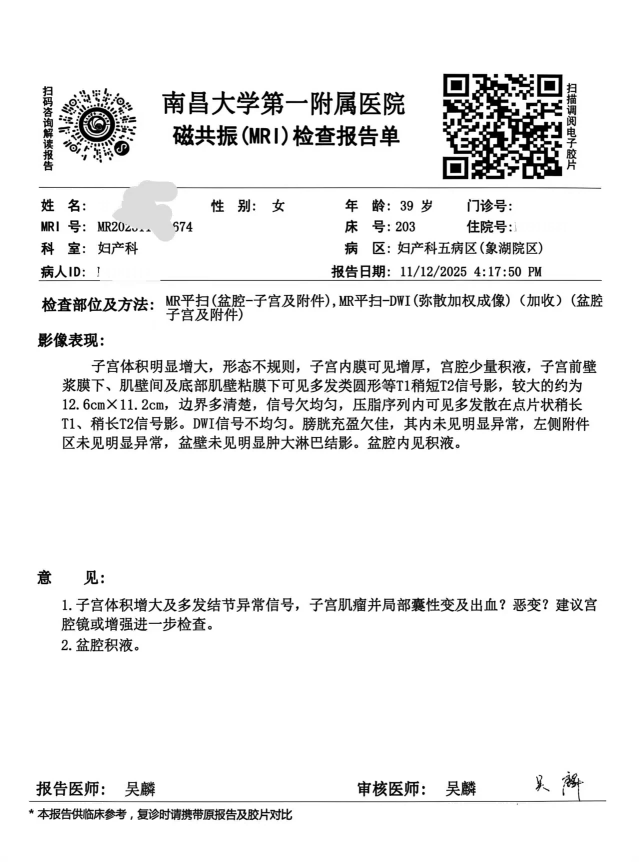
Pelvic MRI Report 盆腔MRI报告
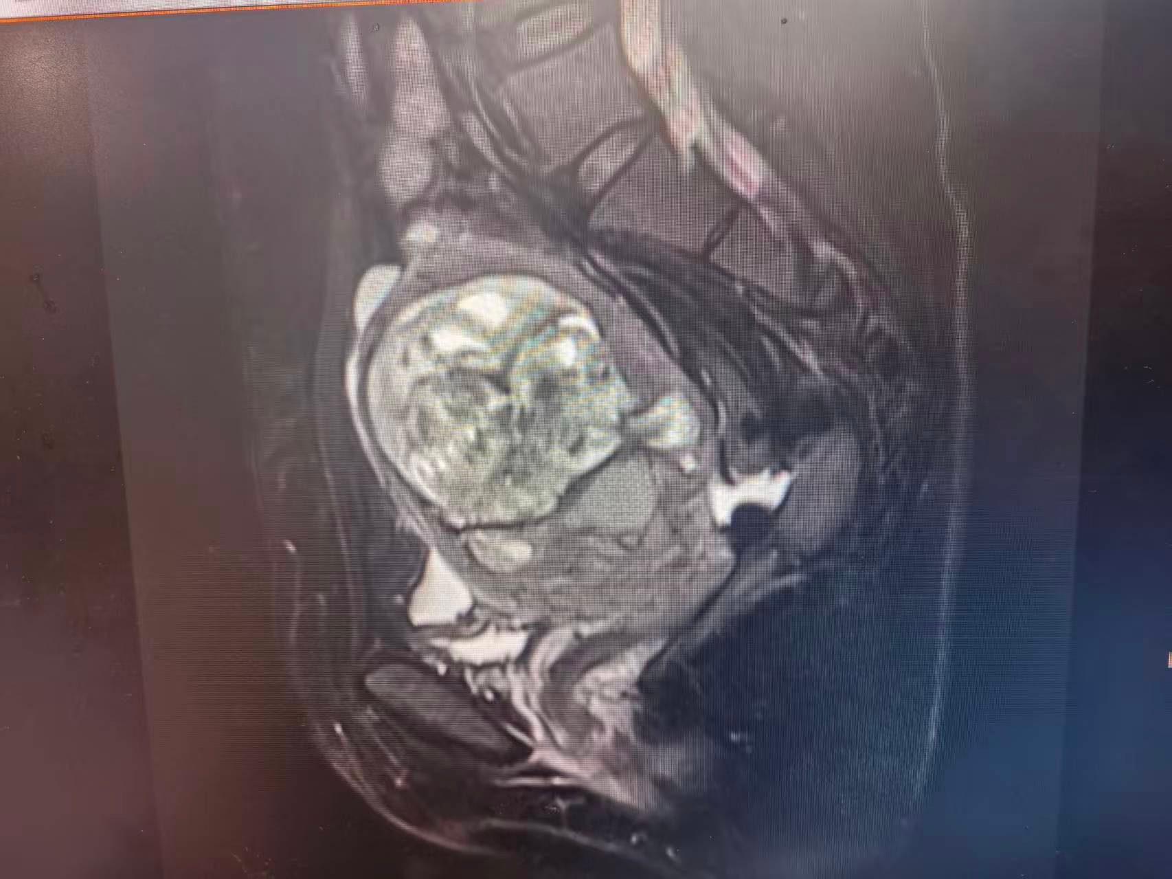
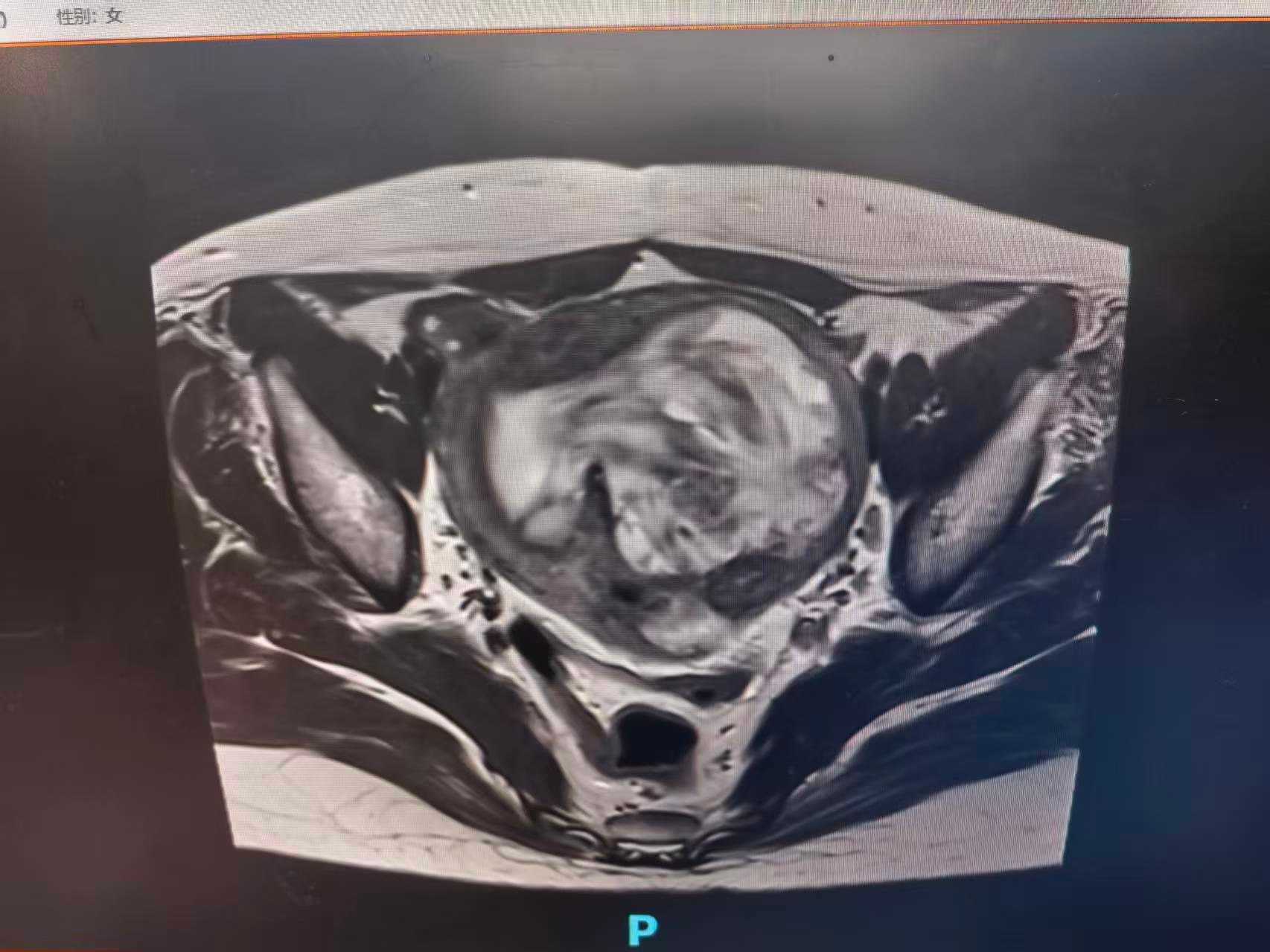
Pelvic MRI Images 盆腔MRI图像
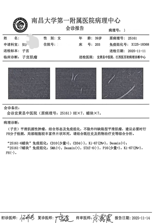
Preoperative Pathological Consultation Results
Preoperative Diagnosis:
Recurrent uterine leiomyoma
Intraoperative Pathology and Subsequent Management:
Intraoperative frozen section pathology indicated: (uterine) endometrial stromal tumor, favoring low-grade endometrial stromal sarcoma. After discussion with the family, the surgical scope was expanded intraoperatively to include bilateral salpingo-oophorectomy, pelvic lymph node biopsy, and multiple biopsies of the greater omentum and peritoneum to achieve staging and radical resection.
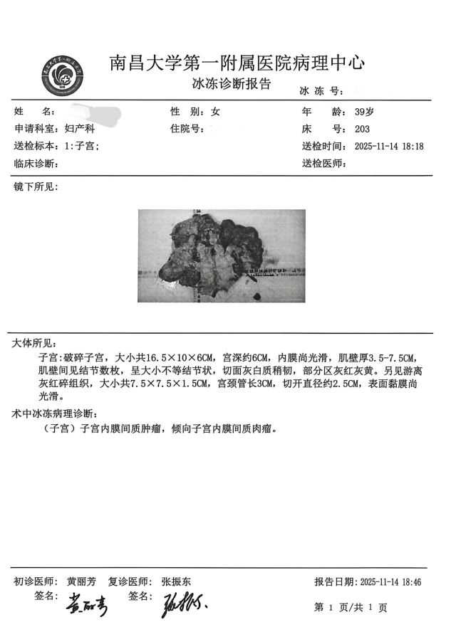
Intraoperative Frozen Section Report
Postoperative Pathological Confirmation:
Low-grade endometrial stromal sarcoma (pT1b) with tumor thrombus identified within lymphatic vessels. No metastasis detected in lymph nodes, greater omentum, or peritoneum. Bilateral adnexa showed benign changes.
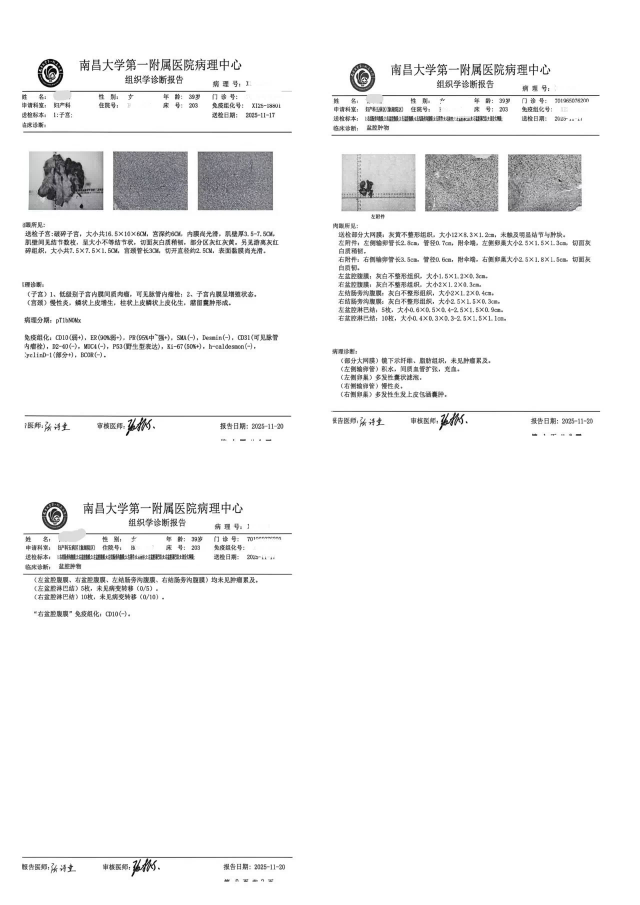
Postoperative Pathology Report
Significance of the Surgery:
This surgical video comprehensively demonstrates how laparoscopic techniques can achieve complete tumor resection and protection of vital organs within a complex pelvic environment characterized by severe adhesions and ureteral anatomical variations. It highlights fine ureteral dissection and ureteric tunnel entry techniques, and we look forward to discussing these approaches with colleagues.
Surgeon Profile
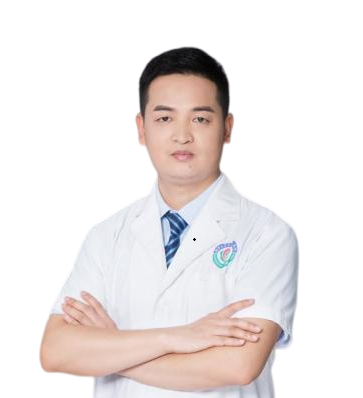
He Quan
Attending Physician, Department of Obstetrics and Gynecology, The First Affiliated Hospital of Nanchang University
Current Positions:
Member, Second Committee of the Obstetrics and Gynecology Branch, Jiangxi Society of Integrated Medicine;
Member, Youth Group, Second Committee of the Gynecologic Oncology Branch, Jiangxi Medical Association;
Member, Second Committee of the Reproductive Health and Family Planning Branch, Jiangxi Medical Association;
Member, Enhanced Recovery After Surgery (ERAS) Branch, Jiangxi Society of Integrated Medicine;
Member, Private Cosmetic Surgery Group, Jiangxi Medical Cosmetology Association;
Member, Brachytherapy Committee, Jiangxi Society of Integrated Medicine.
Academic and Research Achievements:
Principal investigator of one key research project funded by the Jiangxi Provincial Department of Education; participated in multiple provincial- and national-level research projects;
Published several academic papers, with ongoing focus on frontiers in gynecologic oncology and minimally invasive surgery;
Recipient of the Surgical Video Excellence Award at the 10th East China Obstetrics and Gynecology Academic Conference;
Selected for electronic poster presentation of surgical videos at the 2025 Annual Meeting of the Chinese Medical Doctor Association’s Obstetricians and Gynecologists Branch.
Clinical Expertise:
Extensive clinical experience in diagnosing and treating common gynecological conditions and diseases, with long-term dedication to optimizing and applying minimally invasive techniques (laparoscopy, hysteroscopy). Specializes in standardized surgical management and individualized treatment of gynecologic malignancies including cervical, endometrial, and ovarian cancers, while actively promoting Enhanced Recovery After Surgery (ERAS) protocols in gynecology.
Editor: Qinghuan
Reviewer: Maye