Surgical Procedures
- Clamp and pull down the cervix with a cervical forceps.
- Inject diluted epinephrine combined with ropivacaine to create a fluid cushion.
- Make a circumferential incision on the vaginal fornix below the vesicocervical groove.
- Incise the anterior vaginal wall mucosa, then dissect the vesicovaginal space and vesicocervical space.
- Incise the posterior vaginal wall mucosa, then dissect the rectocervical space.
- Clamp the right cardinal ligament, uterosacral ligament as well as uterine arteries and veins, and perform coagulation and transection using LigaSure.
- Suture and ligate the stumps.
- Resect part of the cervix with a scalpel.
- Conduct thorough disinfection.
- Evert the uterus and perform antegrade hysterectomy.
- Clamp the left fallopian tube and round ligament, then achieve coagulation and transection with LigaSure.
- Suture and ligate the stumps.
- Clamp the left utero-ovarian ligament and perform coagulation and transection using LigaSure.
- Suture and ligate the stumps.
- Suture the vaginal angles.
- Perform continuous locking suture on the peritoneum near the vaginal stump using 2-0 absorbable sutures.
- Inject diluted epinephrine combined with ropivacaine to create a fluid cushion again.
- Mark a rectangular area on the anterior vaginal wall: the distal incision is located at the vaginal transverse groove 3 cm below the urethral meatus, and the proximal incision is at the vaginal stump.
- Mark a rectangle of the same size on the posterior vaginal wall corresponding to the tissue to be resected on the anterior vaginal wall.
- Resect the entire fascia of the anterior vaginal wall from top to bottom. Preserve as much fascia anterior to the bladder as possible to strengthen the fascia after suturing.
- Resect the entire fascia of the posterior vaginal wall from top to bottom. Preserve as much fascia anterior to the rectum as possible to strengthen the fascia after suturing.
- Perform continuous purse-string suture on the submucosal fascia of the vaginal wall layer by layer from the inside out using 3-0 PDS sutures.
- Perform continuous locking suture on the mucosal stumps of the anterior and posterior vaginal walls using 2-0 absorbable sutures to completely close the vaginal cavity.
- Inject diluted epinephrine combined with ropivacaine to create a fluid cushion.
- Dissect the scar tissue between the posterior vaginal wall and anterior rectal wall.
- Excise the old scar tissue to form fresh wound surfaces.
- Perform interrupted full-thickness suture on the rectum using 3-0 absorbable sutures to ensure good mucosal apposition.
- Reinforce the rectal muscular layer with horizontal mattress inverting sutures using 3-0 absorbable sutures.
- Dissect the space between the skin and external anal sphincter with tissue scissors.
- Clamp the broken ends of the external anal sphincter with tissue forceps.
- Lift the external anal sphincter with tissue forceps. Insert the index finger into the anus to assess the tension of the sphincter.
- Perform end-to-end suture on the broken ends of the external anal sphincter using 2-0 absorbable sutures, ensuring the external anal sphincter encircles the anal canal.
- Suture the levator ani muscle as well as the superficial and deep transverse perineal muscles using 2-0 absorbable sutures.
- Perform interrupted everting mattress suture on the perineal skin using 3-0 absorbable sutures.
- Perform continuous locking suture on the posterior vaginal wall mucosa using 2-0 absorbable sutures.
- Perform continuous locking suture on the perineal skin using 3-0 absorbable sutures to restore the normal anatomical structure of the perineum.
- Conduct digital rectal examination with the index finger to test the local tension of the reconstructed anal sphincter. The standard is that the anus can just admit one index finger with moderate tension.
- Display the immediate postoperative effect.
Display the surgical effect one month after the operation.
Surgical Highlights and Difficulties
Surgical Highlights
- Fluid separation with diluted epinephrine and ropivacaine is applied intraoperatively, which can reduce intraoperative bleeding and relieve postoperative pain.
- Antegrade hysterectomy is performed after everting the uterus, which facilitates the management of the fallopian tubes and utero-ovarian ligaments at the uterine cornua.
- The vaginal wall is completely resected by dividing the tissue into four quadrants (anterior wall, posterior wall, left and right lateral walls). Meanwhile, maximum preservation of the submucosal fascia of the vaginal wall helps enhance the fascia strength after suturing.
- Purse-string suture is adopted to approximate the fascia of the anterior, posterior and lateral vaginal walls until the vagina is fully closed.
- Concurrent perineorrhaphy during colpocleisis can further reinforce the perineal body, narrow the vaginal introitus and strengthen vaginal closure, thereby reducing the recurrence of postoperative pelvic organ prolapse.
- The repair of grade IV old perineal laceration restores the structure and function of the anal sphincter and relieves the patient from fecal incontinence.
- Locate the broken ends of the bilateral external anal sphincter in the radiating skin folds. Dissect the space between the skin and external anal sphincter with tissue scissors. The operator inserts the index finger into the anus to assess the tension of the lifted external anal sphincter held by tissue forceps, so as to confirm accurate localization of the sphincter.
- End-to-end suture of the broken ends of the external anal sphincter with 2-0 absorbable sutures ensures that the external anal sphincter surrounds the anal canal normally.
Surgical Difficulties
- During the dissection of the anterior and posterior vaginal walls for total colpocleisis, it is necessary to avoid excessive resection of the submucosal fascia while controlling bleeding strictly.
- Dense scar tissue and blurred tissue planes are present in grade IV old perineal laceration. Locating, dissecting and accurately approximating the broken ends of the anal sphincter pose great challenges.
The suture of the anal sphincter broken ends requires tension-free or low-tension repair to prevent sphincter rupture and functional impairment. Surgeons must be familiar with local anatomy to avoid injuries to the bladder, rectum and ureter.
Medical History
The patient is a 79-year-old widowed female.
Chief Complaint
Involuntary defecation for 50 years after delivery, vaginal mass prolapse for 10 years with aggravation for half a year.
Present History
The patient developed involuntary leakage of loose stools after vaginal delivery at home 50 years ago. Ten years ago, she noticed a vaginal mass that protruded while standing and retracted spontaneously in the supine position, accompanied by a bearing-down sensation in the anus, but she did not seek medical treatment. In the past six months, the prolapsed mass deteriorated to the size of a fist; it failed to retract spontaneously in the supine position and would prolapse again immediately after manual reduction. The patient suffered pain while walking and sitting, accompanied by vaginal bloody discharge requiring sanitary pads. She was admitted to our hospital. Physical examination revealed grade IV old perineal laceration and complete uterovaginal prolapse. Surgical treatment was recommended, and the patient was hospitalized for surgery. During the course of illness, the patient had good mental status, normal appetite and sleep. She could not control defecation voluntarily and needed manual assistance for urination. No significant weight change was noted.
Past History
The patient had been in good health previously. She denied chronic diseases such as hypertension, diabetes mellitus, heart disease and nephropathy, as well as exposure to infectious diseases including hepatitis, tuberculosis and typhoid. She had no history of surgery, trauma or blood transfusion, and no known drug or food allergies. She completed regular vaccinations.
Menstrual History
Natural menopause for 30 years.
Marital and Reproductive History
Widowed, with no sexual activity for 20 years.Gravida 5, Para 5, all full-term vaginal deliveries.
Specialized Gynecological Examination
- Vulva: Normal development.
- Vagina: Severe prolapse of anterior and posterior vaginal walls.
- Cervix: Severe cervical erosion with ulcers and chronic inflammatory proliferative tissues on the surface.
- Uterus: Atrophic, no tenderness.
- Adnexa: Bilateral adnexa were atrophic with no palpable abnormalities.
- Anus: Grade IV old perineal laceration with everted rectal mucosa and a rectal laceration of approximately 3 cm. The skin folds from the 9 o'clock to 3 o'clock position around the anus disappeared, and the function of the external anal sphincter was lost.
POP-Q Measurements (Unit: cm)
Aa (+3); Ba (+10); C (+10)Gh (6); Pb (0); TVL (10)Ap (+3); Bp (+8); D (+8)
Auxiliary Examinations
- Gynecological Ultrasound: Retroverted uterus with endometrial thickness of 3 mm. The uterine morphology was basically normal. Bilateral adnexa were obviously atrophic with no abnormal echoes detected.
- Pelvic Floor Ultrasound: Severe cystocele (Greentype II), uterine prolapse. The levator ani hiatus dilated during maximum Valsalva maneuver.
- HPV Test: Negative.
- TCT: Negative for intraepithelial lesions or malignancy (NILM).
Preliminary Diagnoses
- Complete uterovaginal prolapse (Grade IV uterine prolapse)
- Grade IV anterior vaginal wall prolapse (cystocele)
- Grade IV posterior vaginal wall prolapse
- Grade IV old perineal laceration
Surgical Procedure
Vaginal hysterectomy combined with total colpocleisis and repair of grade IV old perineal laceration
Operation Date
April 21, 2025
Introduction to the Surgeon
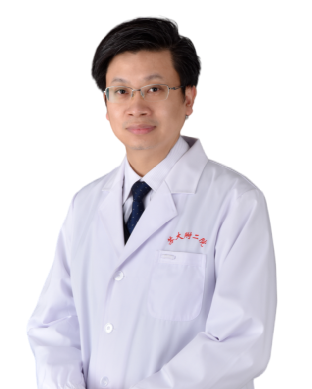
Guo Liangsheng
Assistant Director of the Department of Obstetrics and Gynecology, the Second Affiliated Hospital of Soochow University; Doctor of Medicine; Chief Physician; Associate Professor; Master's Supervisor.
He studied gynecologic oncology, female genital plastic surgery, surgical treatment for uterine prolapse and stress urinary incontinence at Johanniter-Krankenhaus Bonn, Germany in 2017.
Academic Appointments
Young Committee Member of the Gynecologic Oncology Committee, Chinese Association for Research-Oriented Hospitals.
Specialties
Minimally invasive surgery for gynecologic tumors, female genital reconstructive surgery, surgeries for uterine prolapse and stress urinary incontinence.
Academic Honors & Research Achievements
- Academic Titles: Young Key Medical Talent of Jiangsu Province; Outstanding Young Medical Talent of Suzhou City.
- Publications: More than 10 academic papers published in core Chinese medical journals and SCI-indexed journals.
Research Awards: Second Prize of Medical New Technology Introduction Award of Jiangsu Provincial Health Department; First Prize of Suzhou Science and Technology Progress Award; First Prize of Suzhou Medical New Technology Award; Second Prize of Suzhou Medical Science and Technology Award; Second Prize and Third Prize of Jiangsu Maternal and Child Health New Technology Introduction Award.
Editor: Qing HuanReviewer: Ma Ye