Key Surgical Points
- The patient is a young female with no sexual history and obesity, with a BMI of 29.2 kg/m². Pelvic space-occupying lesion was suspected to be a borderline tumor. Single-port laparoscopy was adopted for convenient specimen removal. An exophytic tumor at Stage IC was found intraoperatively. To reduce the risks of recurrence and tumor implantation, intraoperative hyperthermic intraperitoneal chemotherapy (HIPEC) was planned. Drainage orifices were fully utilized to lower surgical difficulty, followed by combined single-port plus auxiliary ports surgery.
- Strict adherence to the no-touch oncologic principle during surgery: All instruments that have come into contact with the tumor, including ultrasonic scalpels, must be replaced.
- Subsequent treatment regimens are formulated based on intraoperative frozen section results and clinical guidelines. For borderline lesions: adnexectomy is performed; for peritoneal biopsy specimens: laparotomy conversion will be conducted if extra-pelvic invasion is detected; for mucinous tumors: appendectomy and omentectomy are required.
Priority is given to fertility preservation and prevention of postoperative abdominal adhesions.
Medical History
Auxiliary Examinations
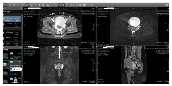
- June 25 (Plain and Contrast-enhanced Pelvic MRI): A pelvic space-occupying lesion measuring 108 mm × 70 mm × 85 mm, highly suggestive of right ovarian cystadenoma with borderline potential. A small amount of pelvic effusion was noted.
- June 25 (Transabdominal Ultrasound): Solid-cystic mass of the right ovary, suspected to be an epithelial-derived borderline ovarian tumor, with a small amount of pelvic effusion.
- June 25 (Tumor Markers): CA199: 52.1 U/ml; other indicators were within normal range (CA125: 45.4 U/ml, HE4: 44.49 pmol/L; premenopausal ROMA index: 6.13%).
Preoperative imaging:
Surgical Procedure
On June 27, the patient underwent laparoscopic ovarian tumor staging surgery under general anesthesia, including right adnexectomy, omentectomy, appendectomy, multiple-site peritoneal biopsy, left ovarian biopsy and left ovarian cyst enucleation. Intraoperative blood loss was minimal.
Intraoperative Frozen Section Pathology Results
- Vegetations on the surface of the right ovary: Serous tumor with borderline changes.
- Right ovary: Borderline seromucinous tumor. Due to limited sampling in frozen sections, further lesions need to be confirmed by routine pathological examination and immunohistochemistry.
- Biopsy specimen of the left ovary: Serous cystadenoma.
- Left ovarian cyst: Serous cystadenoma.Multiple biopsies including vegetations at the root of the mesentery all revealed a small amount of fibrofatty tissue.
Postoperative Conditions
Postoperative Pathological Diagnosis
- Right ovary: Borderline seromucinous tumor.
- Right fallopian tube: Normal tubal tissue.
- Left ovarian cyst & left ovarian biopsy specimen: Serous cystadenoma.
- Biopsy specimens from the left paracolic gutter, right paracolic gutter and rectouterine pouch peritoneum, as well as vegetations at the root of the mesentery: Fibrofatty tissue.
- Appendix and omentum: Fibrofatty tissue.
- No tumor cells were detected in abdominal lavage fluid.
One month postoperatively, the patient had no obvious discomfort. Re-examination of blood routine, biochemical indices and coagulation function showed normal results. The patient had her first menstrual period on July 23, with a 4-day duration and normal menstrual volume, color and texture. Regular follow-up every 3 months was recommended, and no abnormalities have been found so far.
Postoperative incision:

Introduction to the Surgeon

Jiang Jun
- Young Committee Member, Gynecologic Oncology Branch of Zhejiang Medical Association
- Member of Vaginal Pelvic Floor Reconstruction Group, Pelvic Floor Diseases Special Committee of Zhejiang Medical Doctor Association
- Committee Member, Integrated Traditional Chinese and Western Medicine Reproductive Health Special Committee of Zhejiang Society for Mathematical Medicine
- Young Committee Member, Pelvic Integrated Medicine Special Committee of Zhejiang Society for Mathematical Medicine
- Committee Member, Traditional Chinese Medicine Special Committee of Zhejiang Sexology Association
- Committee Member, Gynecological Preventive Treatment Special Committee of Zhejiang Preventive Medicine Association
- Committee Member, Gynecological Urology and Pelvic Floor Reconstruction Special Committee of Zhejiang Senior Scientists Association
- Committee Member, Female Reproductive Health Drug Research Special Committee of China Association of Traditional Chinese Medicine
He has presided over and participated in 8 provincial and ministerial research projects, and published 8 SCI papers.
Editor-in-charge: Qing Huan
Reviewer: Ma Ye