Your Current Location: EXPERT SURGERIES >> 每周一手术 >> Details
88
0
Weekly Surgery | Professor Gong Xiaoming: Lin’s Method Transcervical Resection of Myoma (Type II Fibroids)
In this issue of the "Weekly Surgery" column, we will focus on sharing the wonderful video of "Lin’s Method Transcervical Resection of Myoma (Type II Fibroids)" by Professor Gong Xiaoming, co-founder of Woyi Obstetrics and Gynecology Elite Group and chief expert of Woyi HIFU Center for Uterine Fibroids, providing a reference for everyone to exchange and learn surgical techniques.
妇科手术

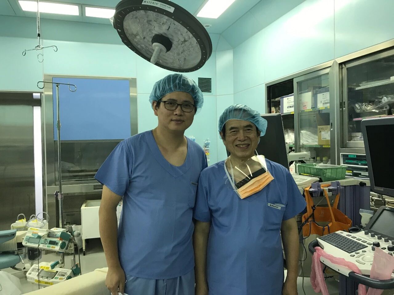
The so-called Lin’s Method was founded by Professor Lin Baoliang, a world-renowned hysteroscopy expert, and is his unique surgical approach for hysteroscopic myomectomy. Professor Lin was born in Taiwan and has lived in Japan for a long time. He has unique research in hysteroscopy technology and has developed many instruments specially for hysteroscopic surgery. In September 2017, after the 18th APAGE (Asia Pacific Association for Gynecologic Endoscopy) Annual Meeting, Professor Gong Xiaoming studied with Professor Lin in Japan for one week, becoming the first doctor from Chinese mainland to learn from him.

微信图片_20260513091509.jpg

Figure caption: Professor Gong Xiaoming (left), Professor Lin Baoliang (right)


The key difference between Lin’s Method TCRM and the traditional TCRM is to find the capsule of the myoma, so the surgical method does not use the resectoscope loop to resect the myoma.


Surgical Steps

Real-time ultrasound monitoring is used during the operation (this is very important), the distension pressure is set at 100 mmHg, and continuous perfusion is maintained in the uterine cavity.
Step 1: Dilate the cervix to No. 12 with a cervical dilator.
Step 2: Use a needle electrode to make a longitudinal incision on the endometrium of the posterior uterine wall to expose the uterine fibroid.
Step 3: Use the resectoscope needle to perform blunt dissection along the capsule of the uterine fibroid to find the space of the pseudocapsule. Dissect deeply enough at the myometrial base near the cervix to expose the space easily. Generally, fibroids have clear boundaries and can be completely separated at the pseudocapsule.
Step 4: After about 3/4 of the myoma is freed, cut the myoma into small pieces with the resectoscope needle, with the needle perpendicular to the edge of the myoma.

Step 5: Dilate the cervix to No. 16. Under the guidance of ultrasound, use Lin’s forceps to clamp and remove the segmented myoma. When clamping the myoma, try to rotate it first to completely free the tumor along the uterine wall.


Case Summary

The patient, 48 years old, married, was admitted due to increased menstrual flow for 2 years. Her menstrual flow increased significantly after a missed abortion 2 years ago, accompanied by increased vaginal discharge, which affected daily life. There was no obvious dysmenorrhea, with a pain score of 0. Regular menstruation, 28-day cycle, 6-day duration, heavy flow, last menstrual period June 1, 2025. Gravida 2, para 0, 2 artificial abortions, no current fertility needs. Previous meniscus repair surgery, no chronic medical history, blood transfusion history, drug allergy history, no family history of tumors; received 9-valent HPV vaccine, no re-examination of HPV and TCT.


Auxiliary Examinations

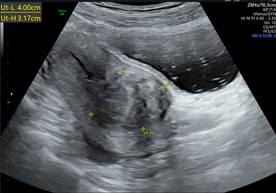
  1. Transvaginal three-dimensional ultrasound on May 21, 2025: Anteverted uterus, uneven myometrial echo, a hypoechoic mass of about 20mm*14mm*18mm at the posterior wall of the uterine fundus, a hypoechoic mass of about 56mm39mm48mm at the right anterior wall of the uterine body, compressing the uterine cavity; cystic structures in bilateral ovaries; suggesting uterine fibroids (one may compress the uterine cavity) and cystic structures in bilateral ovaries.
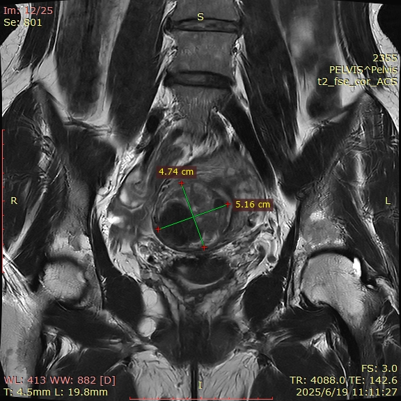
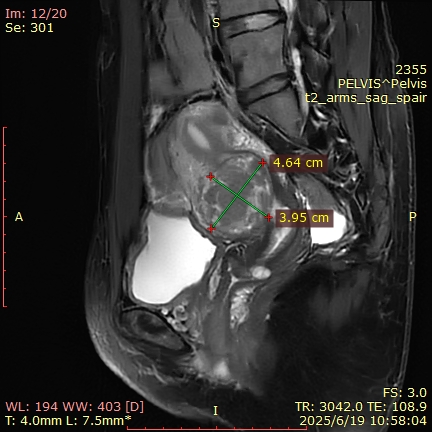
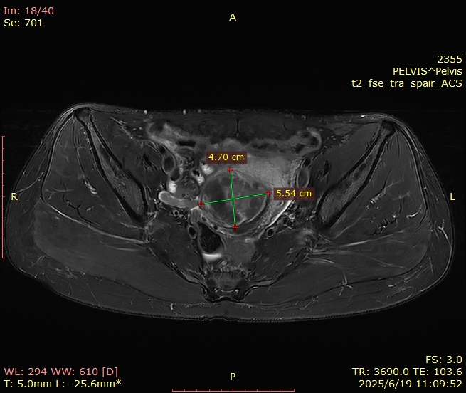
  2. Pelvic MRI on June 19, 2025: Multiple fibroids in the uterine fundus and body, the largest about 50mm*52mm*50mm, protruding into the uterine cavity; multiple cervical cysts; no abnormalities in bilateral adnexa; a small amount of pelvic effusion, left Bartholin’s cyst, sacral canal cyst.
图片1.png

图片2.png

图片3.png


  1. Post-medication ultrasound review: After 4 months and 8 months of Zoladex treatment, the uterus and fibroids gradually shrank. The largest fibroid shrank from the initial 56mm to about 34mm*29mm*26mm, the endometrium became thinner, and there was a small amount of separation in the uterine cavity.

Diagnosis and Treatment Process

  • Oral tranexamic acid was given for symptomatic treatment, and menstrual flow increased and menstrual period prolonged after drug withdrawal.
  • Starting from June 2025, 10.8mg Zoladex was injected subcutaneously, combined with Remifemin and calcium supplements. Menopausal symptoms occurred during treatment and gradually relieved with treatment.
  • After medical pretreatment, the fibroids shrank significantly to 40mm32mm31mm. Hysteroscopic myomectomy (TCRM) was planned. Misoprostol was orally administered for 3 days and cervical dilators were used to prepare the cervix before surgery.

Diagnosis

  1. Uterine fibroids
  2. Menorrhagia


图片4.png

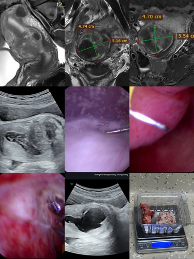
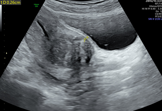
Preoperative uterine fibroid size: 40mm*32mm

图片5.png

Distance from the outer capsule of the uterine fibroid to the serosa layer (SMT): 2.6mm
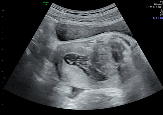

图片6.png

Intraoperative ultrasound imaging showing the location of the uterine fibroid
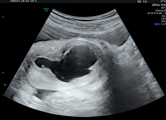

图片7.png

Postoperative cavity imaging after hysteroscopic myomectomy
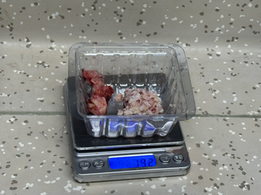

图片8.png

Weight of the resected uterine fibroid specimen: 19.2g


图片9.png


Surgeon Profile

龚老师净版.png

Gong Xiaoming, MD

Chief Expert of Woyi HIFU Center for Uterine Fibroids

Independent practicing obstetrician and gynecologist, founder and editor-in-chief of China Obstetrics and Gynecology Network, co-founder of Woyi Obstetrics and Gynecology Elite Group, former associate professor and master’s supervisor of Obstetrics and Gynecology Department, Peking Union Medical College Hospital, former deputy director of Gynecology Department and director of Minimally Invasive Center, Shanghai First Maternity and Infant Hospital.

Social Part-time Positions

Member and Secretary of Perinatal Health Care Group, Women’s Health Care Branch, Chinese Preventive Medicine AssociationExecutive Director of Female Genital Reconstruction Branch, Chinese Association of Plastics and AestheticsStanding Member of Gynecological Urology Branch, Beijing Medical AssociationMember of Expert Advisory Committee of Maternal and Child Health Industry Branch, National Health Industry Enterprise Management AssociationInternational Member of Patient Education Committee, International Urogynecological Association (IUGA)Director of Innovation Application Council, Internet SocietyExecutive Member of Mobile Medical Professional Committee, Chinese Research Hospital AssociationMember of Maternal and Child Minimally Invasive Professional Committee, Chinese Maternal and Child Health AssociationYouth Member of Medical Promotion Association

Youth Member of Minimally Invasive Non-invasive Committee, Chinese Medical Doctor Association


Editor: Qing HuanReviewer: Ma Ye
评论(0)
精彩评论
Featured Recommendations