Surgical Challenges
Uterine broad ligament leiomyomas are classified into true and false types. True broad ligament leiomyomas arise from myometrial tissue within the broad ligament and have no anatomical connection to the uterus. False broad ligament leiomyomas result from uterine body leiomyomas protruding into the broad ligament.
Their special characteristics are as follows:
1. Complex anatomical relationships: Adjacent to vital structures including the ureter, uterine artery, internal iliac vessels, and obturator nerve. The ureter is often displaced by the tumor and may even adhere to the tumor capsule, making it highly vulnerable to injury.
2. Abundant blood supply: The tumor is mainly supplied by a vascular network within the broad ligament, featuring large, tortuous vessels that are difficult to control, often leading to massive intraoperative bleeding.
3. Limited surgical space: The tumor stretches the anterior and posterior leaves of the broad ligament, forming a “pseudocapsule,” yet the actual working space is narrow with poor exposure.
Single-port laparoscopy, with its single access, restricted space, and instrument interference, further increases surgical difficulty.
Intraoperative Precautions
1. Perform thorough exploration; select the most prominent, least vascular, and ureter-distant area as the entry point. All procedures must be performed only after confirming anatomical relationships.
2. Bipolar electrocoagulation can be used during dissection to reduce bleeding and maintain a clear surgical view.
3. Elevate the tumor to keep it away from the ureter and intestines for safety.
4. Suture the tumor cavity tightly without dead space to prevent hematoma formation.
Case History
Patient Information
· Gender: Female
· Age: 45 years
· Ethnicity: Han
· Marital Status: Married
· Occupation: Unemployed
Chief Complaint
Palpable abdominal mass for 1 month.
History of Present Illness
One month ago, the patient palpitated a firm, non-tender lower abdominal mass about fist-sized while lying flat in the morning. Twenty days ago, she had spontaneous vaginal bleeding of small volume and dark red color, without abdominal pain or distension, no change in menstruation, no vaginal discharge, no frequent urination or defecation difficulty, no weight loss or fatigue. The bleeding stopped spontaneously 3 days ago.Four days ago, ultrasound at a local county hospital suggested uterine leiomyoma and recommended surgery. The patient sought further treatment at our hospital and was admitted with a diagnosis of “uterine leiomyoma.”Since onset, the patient has had normal appetite, sleep, bowel, and bladder function, with no weight loss.
Past Medical History
No history of hepatitis, tuberculosis, kidney disease, hypertension, diabetes, or heart disease. No history of trauma, blood transfusion, or drug/food allergies. Cesarean sections were performed at the local county maternal and child health hospital in 2002 and 2008.
Personal History
No history of traveling to epidemic-stricken areas, no toxic exposure, no smoking or alcohol use.
Marital History
Married at age 24; spouse is healthy.
Menstrual and Obstetric History
Menarche at 12 years old, menstrual period 6–7 days, cycle 25 days, moderate menstrual flow, no dysmenorrhea, last menstrual period June 6, 2024. Gravidity 2, parity 0, abortion 2, living children 2. Contraception: intrauterine device.
Family History
No history of hematological diseases, hypertension, or diabetes in parents. No history of infectious, genetic, cancerous, or tumorous diseases in family members.
Physical Examination
Vital signs: temperature 36.5°C, pulse 92 beats/min, respiration 18 breaths/min, blood pressure 127/89 mmHg.General condition: normal development, good nutrition, natural facial expression, clear consciousness, spontaneous posture.Skin and mucous membranes: normal color, no petechiae or ecchymosis, no edema, scar: transverse scar in lower abdomen about 14 cm long.Superficial lymph nodes: no enlargement.Head and organs: no skull deformity; conjunctiva non-congested and non-pale, sclera non-icteric, pupils equal and reactive to light; ears, nose normal; lips red, pharynx non-congested, tonsils not enlarged.Neck: no resistance, no jugular venous distension, no abnormal carotid pulsation, trachea midline, thyroid not enlarged.Chest: no deformity; symmetric respiratory movement, equal vocal fremitus, resonant percussion, hepatic dullness border at 5th intercostal space on right midclavicular line, clear breath sounds, no pleural friction rub; no precordial bulge, apical impulse at 0.5 cm medial to left midclavicular line in 5th intercostal space, no diffusion, no thrill, no cardiac enlargement, heart rate 92 beats/min, regular rhythm, no murmur.Abdomen: flat, no superficial varicose veins, no hepatosplenomegaly, no muscle tension, no tenderness or rebound tenderness; palpable mass in lower middle abdomen about 4 cm×3 cm, firm; no shifting dullness, no costovertebral angle tenderness, bowel sounds 4 times per minute.Spine and extremities: no deformity, free movement.Nervous system: biceps tendon reflex, abdominal wall reflex, knee tendon reflex normal; Babinski sign, Kernig sign, Brudzinski sign negative.
Gynecological Examination
Vulva: parous.Vagina: patent, non-congested mucosa, small white discharge, no odor.Cervix: normal size, smooth surface, tough, no tenderness on motion.Fornices: empty, no tenderness.Uterus: retroverted, firm mass on left anterior wall about 9 cm×8 cm×7 cm, clear border, good mobility, no tenderness.Adnexa: no obvious abnormality in bilateral adnexal areas.
Auxiliary Examination
Three-dimensional ultrasound, July 8, 2024: Uterus retroverted, uterine body 56 mm×57 mm×47 mm, irregular shape, heterogeneous myometrial echo, left lateral wall exophytic hypoechoic mass 93 mm×92 mm×74 mm (type VII). CDFI: strip blood flow signals inside and around the mass. Endometrial thickness 9 mm. “V-shaped” intrauterine device with normal position. Left ovary not visualized. Right ovary 24 mm×15 mm. Pouch of Douglas effusion 26 mm.
Primary Diagnosis
1. Uterine leiomyoma
2. Intrauterine device
Surgical Procedure
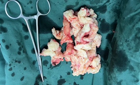
After admission, laboratory examinations were completed. Curettage was performed first to exclude endometrial lesions. Then transumbilical single-port laparoscopic surgery was performed under general anesthesia. A left broad ligament leiomyoma about 9 cm×8 cm×7 cm was found intraoperatively, and myomectomy was performed. The operation was smooth.
Pathology: Uterine leiomyoma with hyaline degeneration.
Surgeon Profile

Sun Xiaohua
Deputy Chief PhysicianDepartment of Gynecologic OncologyQinhuangdao Maternal and Child Health Hospital
Edited by: Qing Huan
Reviewed by: Ma Ye