Case History
A 40-year-old female patient was admitted to our hospital at 20:39 on March 29, 2024, due to "recurrent vaginal bleeding for more than 2 months, 4+ months after cesarean section".
Admission Diagnosis
- Abnormal uterine bleeding
- Placenta accreta with bleeding (after transcervical resection)
- Uterine arteriovenous fistula?
- Scarred uterus (four times)
Uterine diverticulum
Present Medical History
No obvious abnormalities were found in routine blood test, coagulation function test, emergency biochemistry and liver function test after admission, and early pregnancy test was negative.
Marriage and Childbearing History
- 2008: cesarean section of a live baby girl due to "macrosomia", weighing 3300g
- 2013: cesarean section of a live baby girl due to "scarred uterus", weighing 3600g
- 2014: transabdominal embryo removal due to "cesarean scar pregnancy"
- 2016 and 2017: one early pregnancy abortion each
- 2021: spontaneous abortion at 26+ weeks of gestation due to "premature rupture of membranes"
- November 12, 2023: cesarean section at the uterine body, uterine repair and plasty, bilateral ascending branch ligation of uterine artery, bilateral tubal ligation, pelvic adhesiolysis and abdominal adhesiolysis at 28 weeks of gestation due to "central placenta previa with bleeding, placenta accreta with bleeding and scarred uterus", delivering a live baby boy weighing 1325g, currently healthy. Intraoperative blood loss was 3000ml, with transfusion of 8 units of red blood cells and 600ml of plasma.
No special past medical history, menstrual history, personal history or family history.
Physical Examination on Admission
Specialist examination: normal vulva, unobstructed vagina, about 2ml of dark red blood accumulation in the vagina, no active bleeding after wiping. Normal cervical size with Nabothian cysts, no contact bleeding, no cervical motion tenderness. Anteverted uterus, normal size, medium consistency, no tenderness, good mobility. No obvious abnormalities in bilateral adnexa.
Auxiliary Examinations
Admission Tests
- Scarred uterus, no definite signs of residual placenta or arteriovenous fistula, a little blood accumulation in the uterine cavity;
- Cervical Nabothian cysts;
A small amount of effusion in the rectouterine pouch.
Surgical Procedure
On April 2, 2024, the patient underwent laparoscopic total hysterectomy + bilateral salpingectomy + pelvic adhesiolysis + abdominal adhesiolysis + enterolysis + abdominal catheter drainage under combined intravenous-inhalational general anesthesia in the pelvic and abdominal cavity.
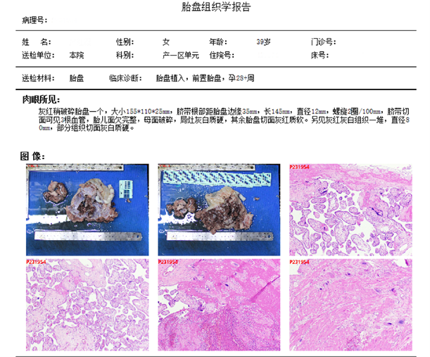
Pathological Diagnosis (I)
- Villous development roughly consistent with gestational age, partial villi with accelerated maturation, increased proportion of terminal villi, focal villous dysplasia; increased fibrinoid deposition with focal calcification under the chorionic plate and around villi, narrow intervillous space in some areas, intervillous thrombosis.
- Umbilical cord with 2 arteries and 1 vein, middle insertion.
- Inflammatory reaction: maternal inflammatory reaction: neutrophil infiltration in decidua and chorionic layer with abscess formation, consistent with stage I severe degree. Fetal inflammatory reaction: no obvious inflammation in umbilical cord and chorionic plate vessels.
- Spiral artery indicators: number of basal plate spiral arteries: 2, remodeling (2), poor remodeling/absent remodeling/fibrinoid necrosis (0). Number of fetal membrane arteries: >10.
- Decidua absence at partial placental implantation site, placental tissue interweaving and closely adhering to uterine smooth muscle tissue, consistent with placenta accreta in combination with clinical findings.
Pathological Diagnosis (II)
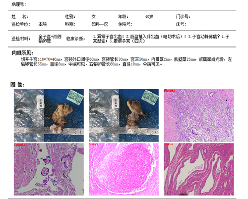
- Chronic cervicitis with multiple Nabothian cysts, partial glandular epithelium with immature squamous metaplasia. Abundant proliferative arteries and veins in cervical stroma with obvious venous lumen dilatation and partial intraluminal thrombosis, consistent with cervical arteriovenous malformation-arteriovenous fistula with focal thrombosis in combination with clinical imaging.
- Adenomyosis with hemorrhage.
- Endometrium with irregular proliferative reaction.
- A very small amount of atrophic villous tissue attached to the local muscular wall surface, consistent with placenta accreta in combination with clinical history.
Chronic inflammation of bilateral fallopian tube mucosa.
Discharge Diagnosis
- Uterine arteriovenous fistula
- Poor postoperative wound healing
- Adenomyosis
- Placenta accreta with bleeding (after transcervical resection)
- Scarred uterus (four times)
Uterine diverticulum
Outpatient Follow-up
Physical examination: vaginal stump wound and abdominal wall wound healed well.
Surgical Difficulties
The difficulty lies in the unclear boundary and severe adhesion between the lower uterine segment, cervix and bladder, making separation extremely difficult and carrying a high risk of bladder injury. Excessive resection of the anterior uterine wall tissue with placenta accreta during the previous cesarean section led to excessive tension and persistent unhealing of the lower uterine segment, resulting in iatrogenic secondary arteriovenous fistula, which was confirmed by pathological sections.
Surgeon Profile

- The first "Most Beautiful Physician in the Eyes of Colleagues"
- The 8th "Young Good Doctor of Yangcheng"
- The 10th Batch of Outstanding Medical Cadres Aiding Xizang in Guangdong Province
- Secretary-General of the Gynecologic Oncology Committee of Guangdong Primary Medical Association
- Standing Member of the Youth Committee of Guangdong Plastic Surgery Association
- Standing Member of the Reproductive Plastic Surgery Branch of Guangdong Plastic Surgery Association
- Member of the Obstetrics and Gynecology Committee of Guangdong Medical Education Association
Member of the Gynecologic Oncology Committee of Guangdong Pharmaceutical Association