Your Current Location: EXPERT SURGERIES >> 手术新秀 >> Details
199
0
Outstanding Young Surgeon | Niu Yuwei: Laparoscopic Resection of Broad Ligament Uterine Fibroids
In this issue of Outstanding Young Surgeon, we will focus on the surgical video of Laparoscopic Resection of Broad Ligament Uterine Fibroids performed by Deputy Chief Physician Niu Yuwei from the Department of Obstetrics and Gynecology, Binhu Campus of Hefei First People's Hospital (The Third Affiliated Hospital of Anhui Medical University).
其他

Treatment Process

After admission, relevant examinations were completed, and there were no surgical contraindications. Laparoscopic resection of broad ligament uterine fibroids was performed under tracheal intubation general anesthesia. Preoperative preparations were fully made, and the patient's physical condition was strictly evaluated to ensure the safe conduct of the operation.

After successful anesthesia, the patient was placed in the lithotomy position. After conventional pneumoperitoneum establishment, a laparoscope was inserted above the umbilical ring. Under the laparoscope, the uterus was full with postoperative changes on the surface; no obvious abnormalities were found in bilateral adnexa. The greater omentum was partially adherent to the anterior uterine wall and right peritoneum. A fibroid-like nodule with a diameter of about 5 cm protruded in the right broad ligament. Further dissection of the broad ligament to free the fibroid revealed that it was located between the internal iliac artery and the right ureter, above the deep uterine vein, growing branching from the cervix along the uterine artery, surrounding and wrapping the ureter and protruding into the broad ligament. Two fibroid-like protrusions with a diameter of about 2 cm and smooth surfaces were seen on the anterior cervical wall.


Surgical Procedures

  1. Separate the adhesions of the surrounding tissues and restore the original anatomical structure.
  2. Cut the right round ligament with a harmonic scalpel, dissect and expose the internal iliac artery, free the initial segment of the uterine artery, and gradually incise the peritoneum around the tumor to free the fibroid. The fibroid-like tissue was lobulated with three branches about 5 cm in size, surrounding the uterus, ureter and uterine artery. The right ureter was freed with a harmonic scalpel, avoiding blood vessels. The blood vessels around the tumor were disconnected at the cervical junction, and the fibroid was completely stripped with precise hemostasis.
  3. Open the capsule of the anterior uterine wall fibroid with unipolar electrocautery, perform sharp and blunt dissection to completely strip the fibroid, and suture and close the tumor cavity with barbed suture. The other fibroid was treated in the same way.
  4. Place the fibroid into a self-made specimen bag, perform morcellation and resection, and remove it together with the specimen bag through the second puncture port. No obvious residual fibroids were found in the pelvic and abdominal cavity.
  5. Perform precise hemostasis with bipolar electrocoagulation, irrigate and inspect the pelvic and abdominal cavity, and confirm no active bleeding on the wound surface.
  6. Evacuate the gas in the abdominal cavity, remove the laparoscope and cannulas, and suture the skin incision. The operation was completed.
The intraoperative anesthesia effect was satisfactory, the patient's vital signs were stable, and the bleeding was minimal. The patient returned to the ward safely after anesthesia recovery.
Specimen: Broad ligament fibroid + uterine fibroid (morcellated), described as above. It was shown to the family members and then sent for pathological examination.

Postoperative Pathology: Leiomyoma.


Case History

Patient, female, 37 years old.

Chief Complaint

Pelvic mass detected on physical examination for more than 2 months.

History of Present Illness

The patient had regular menstruation in the past. Uterine fibroids were found during cesarean section 10 years ago, followed by regular re-examinations. Laparoscopic myomectomy was performed in our hospital in 2020. Transvaginal color Doppler ultrasound re-examination on June 8, 2025 showed multiple hypoechoic lesions in the myometrium, the largest in the anterior wall with a size of 20mm13mm18mm, clear boundary and regular shape, some slightly protruding outside the uterine contour; endometrium was centered with a thickness of 8.8mm. Tortuous strip-like hypoechoic lesions of 50mm19mm and 46mm14mm were detected in the right pelvic cavity with clear boundaries, seemingly connected, and rich blood flow signals were observed inside. Observation and follow-up were recommended. The patient requested admission for surgical treatment.

Past History

The patient was in good health. She underwent cesarean section in 2015 and laparoscopic myomectomy in 2020 with benign pathology.

Menstrual History

Age of menarche: 14 years old, menstrual period 5 days, cycle 29 days. Last menstrual period: August 8, 2025. Regular menstrual cycle, moderate menstrual flow, no dysmenorrhea.

Marital and Childbearing History

Married with one child, G2P1. Her husband and son are healthy.

Family History

No family history of genetic diseases or tumors.

Other Medical History

No special conditions.

Gynecological Examination

Vulva: Normal development, parous.
Vagina: Unobstructed, normal mucosa, no abnormal odor.
Cervix: Smooth, medium consistency.
Uterus: Anteverted, full. A mass of about 5 cm in size was palpable beside the right uterus with unclear texture, no uterine tenderness.
Bilateral adnexal areas: No abnormal masses palpable, no tenderness.

Auxiliary Examinations

Ultrasonography
图片1.png

图片2.png

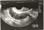
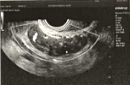
Ultrasonic findings: Uterus anteverted, normal size and regular shape, homogeneous myometrial echo. Multiple hypoechoic lesions in the myometrium, the largest in the anterior wall with a size of 19mm13mm, clear boundary, some slightly protruding outside the uterine contour; endometrial thickness 6mm. Bilateral ovaries are normal in size and shape. Hypoechoic lesions of 55mm17mm and 44mm*15mm with irregular shape and clear boundary are detected in the right pelvic cavity, with slightly rich blood flow signals (RI: 0.53). No obvious mass echo in the left adnexal area. CDFI and PW: No obvious abnormal blood flow signals in the uterus and left adnexal area.
Ultrasonic findings: Uterine fibroids (FIGO type 4, 5); Hypoechoic lesion in the right pelvic cavity (vascular leiomyoma?).

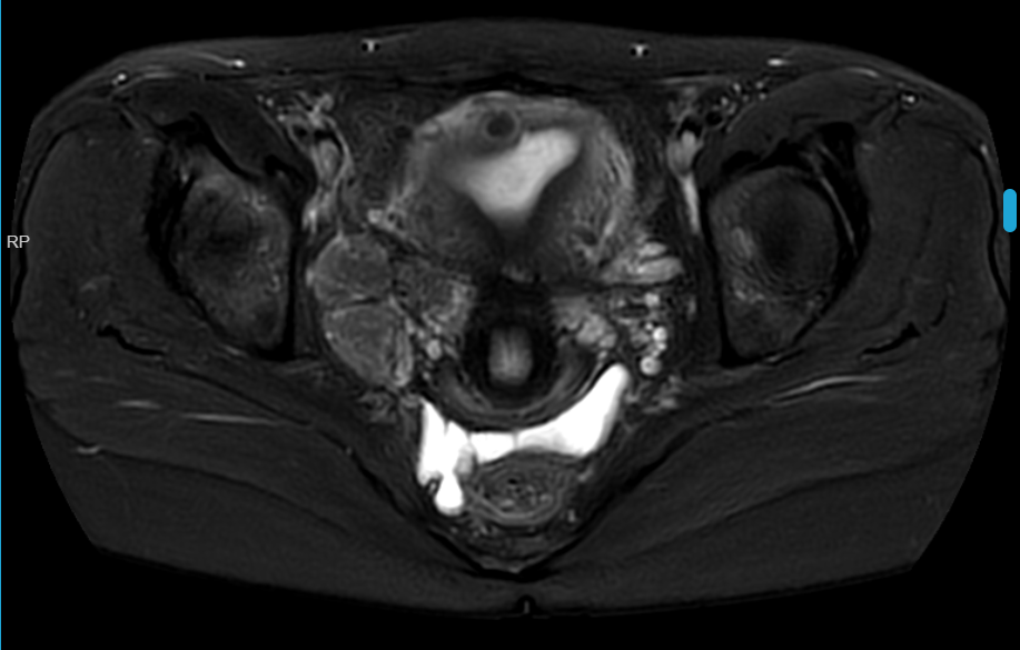
Pelvic Magnetic Resonance Imaging (Plain + Enhanced Scan)
图片3.png

图片5.png

图片4.png

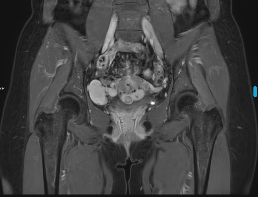
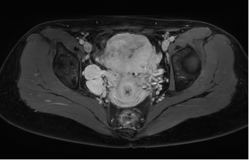
Imaging findings: The uterine surface is uneven. Multiple punctate and nodular lesions with low T1 signal and slightly low T2 signal are seen in the myometrium and under the serosa, with clear boundaries. The largest lesion is about 14.5mm in long diameter with heterogeneous enhancement. Three lesions with low T1 signal and slightly high mixed T2 signal are seen in the right pelvic cavity, with obvious and relatively homogeneous enhancement and smooth edges. The largest lesion is about 23.3mm*38mm.
MRI findings:
  1. Multiple uterine fibroids;
  2. Multiple abnormal signals in the right pelvic cavity, benign tumor suspected, clinical correlation recommended.

Preliminary Diagnosis

  1. Pelvic mass to be diagnosed;
  2. Uterine fibroids;
  3. Scarred uterus.

Discharge and Outcome

The patient recovered well after surgery with good mental state, normal appetite and sleep, no vaginal bleeding, no abdominal pain or distension. Physical examination showed stable vital signs and normal cardiopulmonary auscultation.

Postoperative instructions: rest, avoid heavy physical activities, abstain from sexual intercourse and tub bathing for 1 month. Follow-up visits at the gynecological clinic 1 week and 1 month after surgery.


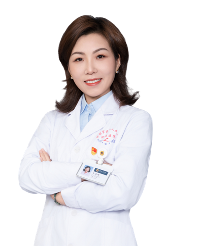
Surgeon Profile
牛玉伟(1).png
Niu Yuwei
Deputy Chief Physician, Member of the Communist Party of China
Department of Obstetrics and Gynecology, Binhu Campus of Hefei First People's Hospital (The Third Affiliated Hospital of Anhui Medical University)
Graduated from Anhui Medical University, engaged in obstetrics and gynecology clinical work for nearly 20 years, studied minimally invasive surgery at the First Affiliated Hospital of Sun Yat-sen University.

Proficient in the diagnosis and treatment of various common, frequently-occurring and critical illnesses in obstetrics and gynecology. Specializes in the diagnosis and treatment of uterine fibroids, adenomyosis, endometriosis, ovarian tumors and other diseases, standardized treatment of gynecological malignant tumors (such as cervical cancer, endometrial cancer and ovarian cancer). Skilled in complex medium and large gynecological surgical skills including vaginal hysterectomy, perineal repair, cervical conization, laparoscopic total hysterectomy for large uterus, myomectomy, fertility-preserving surgery for ectopic pregnancy, pelvic adhesiolysis, ovarian cystectomy, hysteroscopy, endometrial polypectomy, hysteroscopic removal of intrauterine device, hysteroscopic resection of type II submucosal uterine fibroids.

Edited by: Qing Huan
Reviewed by: Ma Ye


评论(0)
精彩评论
Featured Recommendations