Surgical Background
The Mirena intrauterine system (levonorgestrel-releasing intrauterine system) works primarily by locally releasing progestin in the uterine cavity to suppress endometrial hyperplasia, reduce menstrual flow or even induce amenorrhea. It treats dysfunctional uterine bleeding, improves anemia, helps manage adenomyosis and endometrial polyps, prevents endometrial hyperplasia and avoids endometrial cancer, while also providing contraception. It is effective for five years. However, the risk of intrauterine device expulsion or displacement increases when the uterine depth exceeds 9 cm. Therefore, hysteroscopic fixation of the Mirena can better prevent device expulsion and optimize its therapeutic effect.
Hysteroscopic suture fixation of the Mirena uses one stitch of non-absorbable suture to fix the device at the fundus uteri. The surgical challenges include a narrow operating space in the uterine cavity, difficulty with needle-holding and suturing under hysteroscopy, and challenges in inserting and removing the threaded needle through the cervical canal. When instruments are limited (e.g., no hysteroscopic needle holder), suturing can be completed under direct vision with a miniature hysteroscope using a laparoscopic needle holder. The innovation of this procedure is that it effectively resolves Mirena expulsion in enlarged uteri, improves the success rate of uterus-preserving treatment, and helps maintain long-term efficacy of the Mirena.
Various methods of hysteroscopic Mirena fixation are used in China; the procedure presented here is the surgeon’s preferred technique—hysteroscopic suture fixation of the Mirena.
Surgical Procedure
- Hysteroscopy was inserted: normal uterine cavity morphology, uterine depth 10 cm, bilateral tubal ostia visible, slightly thickened endometrium without abnormal blood vessels. Endometrial curettage and biopsy were performed.
- Polyester non-absorbable suture X519H was threaded onto a round needle.
- The needle was inserted into the fundus uteri using a hysteroscopic needle holder.
- Hysteroscopy confirmed the needle was placed at the fundus; the needle holder adjusted the needle direction. The midpoint of the needle was clamped, and one transverse stitch was placed 0.5–1 cm from the fundus on the midline of the posterior uterine wall at a depth of approximately 0.5 cm. The needle holder assisted in needle withdrawal. The threaded needle was removed after successful passage.
- The original Mirena tail string was cut off. The middle section of the 引出 polyester suture was tied around the main body of the Mirena (0.5 cm from the top) for 2–3 loops and secured with knots. The tied Mirena was placed at the fundus using a ring-removing forceps, and the distal polyester suture was gently pulled until taut.
- Hysteroscopy was reinserted to confirm proper positioning of the Mirena with arms extended to both cornua. Both ends of the polyester suture were knotted 3–4 times with a knot pusher to secure the Mirena. Hysteroscopy verified correct knot placement; the suture was cut 0.5 cm outside the knot with hysteroscopic scissors. Final hysteroscopy confirmed normal position, secure fixation, and no bleeding. The procedure was completed.
Follow-up after surgery showed normal position of the Mirena, significantly reduced menstrual flow, resolved anemia, and satisfactory outcomes.
Medical History
Patient, 40 years old, office worker; increased menstrual flow for 2 years.
History of Present Illness
Increased menstrual flow and dysmenorrhea for 2 years, menstrual period 7–10 days, using 20 night sanitary pads per cycle. Hysteroscopy with curettage was performed at our hospital 1 year ago; pathology showed endometrial hyperplasia. Mirena was inserted subsequently but expelled 3 months later. Menorrhagia persisted. Mirena was reinserted 2 months ago and expelled again 1 month ago. The patient was admitted for hysteroscopic Mirena fixation.
On June 22, 2024, ultrasound showed enlarged uterus with abnormal echo suggestive of adenomyosis. Blood routine: HGB 83 g/L. Hysteroscopy, curettage, and Mirena fixation were recommended. The patient was admitted with diagnoses of adenomyosis, AUB-A, and anemia.
Obstetric history: G2P1, one term vaginal delivery, one abortion.
Gynecological Examination
Vulva: married, parous. Vagina: patent, small amount of white discharge. Cervix: smooth, normal size, no contact bleeding, no cervical motion tenderness. Uterus: anteverted, enlarged to size of 2-month gestation, medium consistency, non-tender. Adnexa: no abnormalities.
Auxiliary Examinations
Ultrasound: enlarged uterus with abnormal echo, suggestive of adenomyosis. Blood routine: HGB 83 g/L.
Diagnosis
Adenomyosis; AUB-A; moderate anemia.
Surgical Information
After thorough informed consent, the patient underwent hysteroscopy, curettage, and Mirena fixation under combined intravenous-inhalation general anesthesia on July 3, 2024. The procedure was smooth and lasted 20 minutes. Postoperative pathology: endometrial hyperplasia.
Follow-up to date: significantly reduced menstrual flow, resolved anemia; ultrasound confirmed normal position of the Mirena.
Surgeon Profile
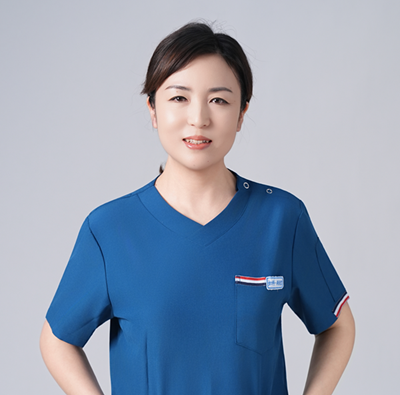
Zhou RunjunMember of the Communist Party of China, Master of Obstetrics and Gynecology, Attending Physician, Department of Gynecology, Tongliang District People’s Hospital, Chongqing; Teaching Secretary, Obstetrics and Gynecology Teaching and Research Office
More than 10 years of clinical and teaching experience in gynecology. Trained in gynecological endoscopic techniques at the National Endoscopy Training Base, Ruikang Hospital Affiliated to Guangxi University of Chinese Medicine; further trained in gynecologic oncology at Beijing Obstetrics and Gynecology Hospital in 2023. Third Prize in the “National Gynecologic Surgery Video Competition” at the 2025 Huaxia Forum on New Gynecologic Diagnosis and Treatment Technologies.
Proficient in minimally invasive gynecologic procedures including family planning operations, hysteroscopy, and laparoscopy. Part-time teacher at Chongqing Medical University and Chongqing Medical College. Awarded Second Prize in Teaching Rounds, Most Popular Teacher in Lectures, and twice honored as Outstanding Teacher at Tongliang District People’s Hospital.
Participated in multiple clinical studies; presided over and completed 1 college-level research project, 1 district-level project, and 2 hospital-level projects; involved in numerous collaborative clinical researches.
Editor-in-Charge: Qing Huan
Reviewed by: Ma Ye