
On March 16, 2026, the pathology team of Jinan Maternal and Child Health Hospital published an article titled "Clinicopathologic and Molecular Landscape of Villitis of Unknown Etiology: Insights From a Large-Scale Case– Control Study in PR China.
The study established a large case-control cohort of VUE from China, and systematically analyzed the clinicopathological correlates and their molecular characteristics of VUE. The article is now compiled as follows:
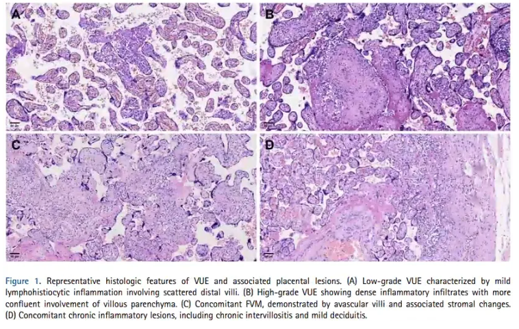
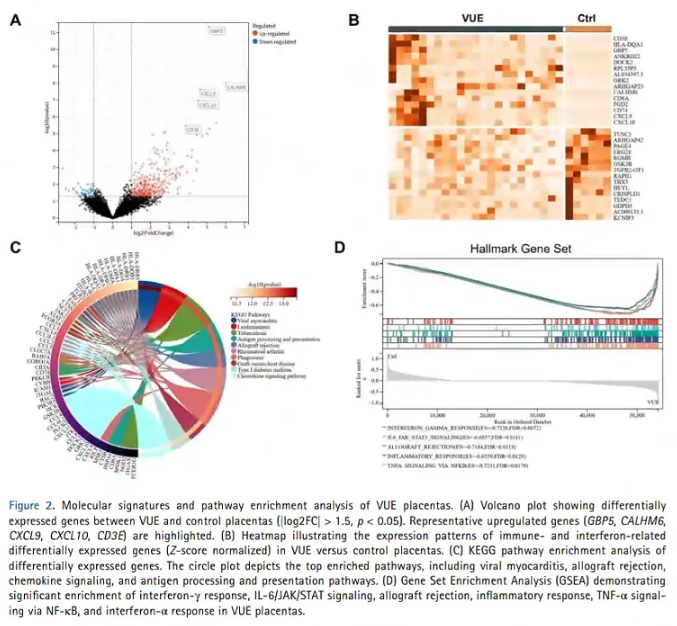
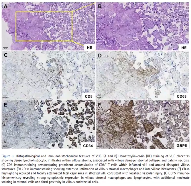
Villitis of unknown etiology (VUE) is a chronic inflammatory lesion of the placenta with no clear basis for infection, mainly manifested by lymphocyte infiltration and destruction of villous structure in the villous interstitium. Although studies have suggested that VUE is associated with fetal growth restriction and adverse pregnancy outcomes, its clinicopathological characteristics and molecular basis are still unclear. This study reviewed and analyzed the VUE cases in the pathology database of Jinan Maternal and Child Health Hospital from 2020 to 2023, and divided them into high and low grades according to Amsterdam criteria. Maternal complications and neonatal outcomes were collected, risk factors were analyzed by multivariate logistic regression, and RNA sequencing was performed on some placental samples. A total of 970 cases of VUE were included, including 381 cases of high-grade and 589 cases of low-grade control, and 980 cases of control, with a total prevalence of VUE of 5.8%. The results showed that hypertensive disorders of pregnancy were independently associated with VUE (OR = 1.67, 95% CI 1.28 to 2.19, p<0.001). VUE placenta was more often accompanied by chronic chorionicitis, chronic decidylitis, and avascular villi, while maternal vascular perfusion was not significantly different from that of controls. High-grade VUE was significantly associated with low birth weight, small for gestational age infants, and increased NICU occupancy. Transcriptome analysis showed that interferon and chemotactic-related genes such as GBP5, CXCL9, and CXCL10 were significantly up-regulated in VUE placenta, and IFN-γ, IL-6/JAK/STAT3, TNF-α/NF-κB and antigen presentation pathways were significantly enriched. Research suggests that high-grade VUE, in particular, is a clinically important placental immune inflammatory lesion whose molecular characteristics support maternal anti-fetal T cell-mediated immune processes. GBP5 may be a potential marker reflecting interferon-driven inflammation. Villitis of unknown etiology (VUE) is one of the common chronic inflammatory lesions of the placenta, which is histologically characterized by villous interstitial lymphocyte infiltration and villous structure destruction, but lacks a clear infectious agent. According to the Amsterdam Consensus, VUE can be divided into low and high levels. The incidence of this disease is about 5%-15% in full-term pregnancies and is thought to be closely related to fetal growth restriction, stillbirth, and recurrent adverse pregnancies. Previous studies have suggested that VUE may be an abnormal maternal immune response against semi-allogeneic fetuses with immunological features similar to transplant rejection. Maternal CD8⁺ T cells and macrophages were commonly infiltrated in the lesion, accompanied by upregulation of CXCL9, CXCL10 and other molecules, suggesting interferon-mediated inflammatory activation. On the other hand, some cases exhibit antiviral-like immune characteristics, suggesting that the mechanism may be heterogeneous. Due to the lack of previous large-sample clinicopathological and molecular integration studies, this study aimed to systematically analyze the clinicopathological characteristics and molecular atlas of VUE. This study has been approved by ethics. Cases diagnosed with VUE were retrospectively searched in the pathology database from 2020 to 2023 and reviewed by pathologists according to Amsterdam criteria. After excluding chromosomal abnormalities, major structural abnormalities and multiple pregnancies, confirmed VUE cases were included. During the same period, the placenta of the single fetus sent for inspection without VUE was used as a control. The placenta was taken from the placenta according to the routine pathological procedure, fixed, paraffin-embedded and sectioned, and the VUE was classified into high and low grades. In order to analyze the molecular characteristics, RNA sequencing and pathway enrichment analysis were performed on some FFPE placenta samples, and GBP5, CD8, CD68 and CD34 immunohistochemistry were further performed in some cases. Statistics were compared between groups and multivariate logistic regression analysis, and the difference was statistically significant with a p<0.05. A total of 48,357 cases were delivered in hospital during the study period, of which 16,267 were sent for testing of placenta singleton. A total of 970 cases of VUE were diagnosed, including 381 cases of high-grade disease and 589 cases of low-grade grade, and the total prevalence of VUE in singleton pregnancies was 5.8%. In addition, 980 cases of placenta singletonis were selected as controls. There were no significant differences in maternal age and gestational age between the three groups. Compared with the control, the incidence of gestational hypertension, gestational diabetes mellitus, and scarred uterus was significantly increased in the high-grade VUE group. Further multivariate analysis showed that only gestational hypertension was independently associated with VUE after correction (OR=1.67, 95% CI 1.28–2.19, p<0.001), suggesting that maternal hypertension may be an important risk factor for VUE. In terms of placental pathology, VUE is closely related to a variety of chronic placental inflammation and fetal circulatory abnormalities. Compared with the control, chronic villous space inflammation and chronic decihyptitis were significantly increased in VUE placenta. The incidence of avascular villus was also significantly increased, with 7.8% in the control group, 16.8% in the low-grade VUE group, and 30.7% in the high-grade VUE group. On the contrary, there was no significant difference in maternal vascular perfusion and acute inflammatory lesions between the three groups. This suggests that VUE, especially high-grade VUE, is more likely to be accompanied by chronic inflammation and impaired fetal circulation. In terms of neonatal outcomes, the average birth weight of infants in the high-grade VUE group was lower, the NICU occupancy rate was higher, and the incidence of low-birth weight infants and small-gestational age infants was also significantly higher than that of the low-grade VUE group and the control group, indicating that the effect of VUE on perinatal outcomes was related to the severity of lesions. There was no significant difference in neonatal hyperbilirubinemia and pneumonia between the three groups. At the molecular level, RNA transcriptome sequencing results showed that a total of 338 genes were up-regulated and 48 genes were down-regulated in VUE. Among them, GBP5 was the most significantly regulated, and inflammation-related genes such as CXCL9 and CXCL10 were also significantly increased. Immunohistochemistry further confirmed that GBP5 was expressed in strong cytoplasm in VUE villous mesenchymal macrophages and lymphocytes, and was expressed in some stromal cells and focal villous endothelial cells, suggesting that it may be a lesion-related marker. HE and CD8, CD68, and CD34 staining also showed cytotoxic T cell and macrophage aggregation in VUE lesions, as well as fetal capillary reduction, suggesting local vascular damage. Pathway enrichment analysis showed that up-regulated genes were mainly enriched in immune regulation, antigen processing and presentation, cytokine-receptor interaction, and angiogenesis. GSEA further showed significant activation of IFN-γ, IL-6/JAK/STAT3, TNF-α/NF-κB, graft rejection, and IFN-α pathways. Overall evidence supports that VUE is a placental immune damage characterized by interferon signaling and T cell driving inflammation. This study combined large-sample clinicopathological analysis with transcriptomics, suggesting that VUE is a maternal T cell-mediated placental inflammatory lesion with clear clinical consequences. One of its most important clinical links is its independent association with hypertensive disorders of pregnancy. The authors suggest that this link may be related to systemic inflammation, placental stress, and endothelial dysfunction in hypertensive pregnancy, but the relationship may also be bidirectional: hypertension promotes VUE, or the inflammatory environment formed by VUE in turn exacerbates maternal hypertension, or the two share upstream maternal-fetal immune tolerance disorders. Since this study is a retrospective design, causal inferences cannot be made. VUE was significantly associated with chronic chorionicitis, chronic decidylitis, and avascular villi without significantly increased maternal vascular malperfusion or acute inflammatory changes. This suggests that VUE is closer to a chronic, sterile, adaptive immune-mediated lesion than an acute infectious process. In particular, in high-grade VUE, there was a significant increase in avascular villi, suggesting a strong link between villous inflammation and impaired fetal circulation, which may also constitute an important bridge between VUE leading to fetal growth restriction and adverse perinatal outcomes. In terms of perinatal outcomes, high-grade VUE was closely associated with low birth weight, small for gestational age infants, and increased NICU occupancy, indicating that the more severe the lesion, the more obvious the clinical impact. This suggests that in routine placental pathology reports, not only the presence of VUE should be reported, but also graded and concomitant vascular lesions should be indicated as much as possible to improve perinatal risk stratification and pertinence in neonatal management. The molecular results further strengthen the immunological interpretation of VUE. GBP5, CXCL9 and CXCL10 and other interferon and chemotaxis-related genes were significantly up-regulated, and IFN-γ, IL-6/JAK/STAT3, TNF-α/NF-κB and graft rejection pathways were active, combined with CD8⁺ T cells and macrophage infiltration, supporting VUE is not a simple morphological diagnosis, but a placental injury state with clear interferon enrichment and cytotoxic immune characteristics. GBP5 is the most prominent molecule in this study and has been immunohistochemistically validated, suggesting that it is a potential marker of interferon-driven inflammation in VUE. The authors also point out that VUE may not be a single mechanism disease, but a group of lesions in which the same immune response overlaps with the antivirus-like immune response. The advantages of this study are that the number of cases is large, the pathological diagnosis has been standardized review, and clinical, histological, and transcriptome information is integrated. The limitation is that only the placenta sent for examination is analyzed, and there may be selection bias; The study was a retrospective design; In addition, there is a lack of maternal-fetal HLA typing and T cell receptor profiling, which cannot directly verify the mechanism of alloimmune immunity. In general, this study systematically depicts the clinicopathological and molecular profile of VUE, indicating that VUE, especially high-grade lesions, has important clinical and biological significance. Compilation Wang Meiling Junior laboratory technician, master of immunology His main research interests are macrophage function Audit Li Juan Chief physician, director of the Department of Pathology, Jinan Maternal and Child Health Hospital Affiliated to Shandong First Medical University Master of Obstetrics and Gynecology from Shandong University; He studied in the Department of Pathology, UPMCMagee Maternity Hospital, Pittsburgh, USA. Academic part-time: member of the Chinese Colposcopy-Pathology Branch (CSCCP), member of the Pathology Professional Committee of the Chinese Maternal and Child Health Association, member of the Pathology Branch of the Shandong Medical Association, vice chairman of the Pathology Branch of the Shandong Maternal and Child Health Association, standing member of the Standing Committee of the Shandong Preventive Medicine Association, and vice chairman of the Pathology Branch of the Jinan Medical Association; He has participated in national, provincial and municipal pathological quality control, and served as a member of the Shandong Provincial Pathological Quality Control Expert Committee and the vice chairman of the Jinan Pathological Quality Control Expert Committee.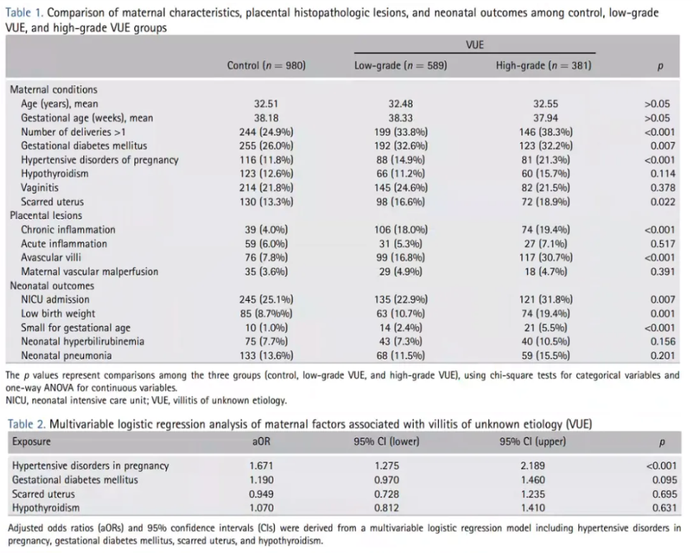


Editor-in-charge:lucy