





















Pregnancy complicated by thoracic aortic disease is a critical condition that seriously threatens the lives of both mother and fetus. Currently, there is a lack of unified clinical diagnostic and therapeutic standards in China. To fill this gap, the Thoracic Aorta Committee of the Chinese Medical Doctor Association Society of Cardiovascular Surgery, the Maternal-Fetal Critical Care Committee of the China Health Promotion Association for Science and Technology of Human Health, and the Cardiac Surgery Branch of the Beijing Medical Association have jointly formulated the Expert Consensus on the Diagnosis and Treatment of Pregnancy Complicated by Thoracic Aortic Disease (2026), integrating national conditions with domestic and international research progress. This consensus addresses eight key clinical issues, including diagnosis and assessment, treatment principles, and pregnancy guidance, forming a total of 45 recommendations (see Table 1).
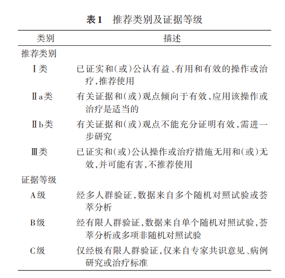
Table 1. Overview of Recommendations
I. The Impact of Pregnancy on the Aorta
To meet the demands of uteroplacental circulation, significant hemodynamic adaptations occur during pregnancy:
(1) Increased Aortic Wall Tension: Cardiac output increases by 30%–50%, accompanied by an increased heart rate (20%–40%) and stroke volume. Peripheral vascular resistance decreases before 20 weeks of gestation, leading to a physiological drop in blood pressure; after 20 weeks, blood pressure gradually rises, increasing by 10%–20% compared to pre-pregnancy levels until delivery. Circulating blood volume peaks at 32–34 weeks of gestation, subsequently increasing aortic wall tension and causing a physiological increase in aortic diameter of approximately 5%. In pregnant women with Marfan syndrome (MFS), aortic dilation is more pronounced, reaching approximately 23%.
(2) Microstructural Remodeling of the Aortic Wall: Changes in estrogen and progesterone levels during pregnancy can induce microstructural changes in the aortic media and intima, manifesting as fragmentation of reticular fibers, loss of the wavy structure of elastic fibers, and reduced glycosaminoglycan content, thereby reducing the aorta's tolerance to hemodynamic load. Animal experiments suggest that oxytocin is closely associated with the occurrence of aortic dissection (AD) during late pregnancy and within 4 weeks postpartum. Additionally, the enlarged uterus compresses the iliac arteries, leading to increased downstream flow resistance.
Consequently, these physiological changes during pregnancy may elevate the risk of thoracic aortic pathology.
II. Multidisciplinary Team (MDT) for Pregnancy Complicated by Thoracic Aortic Disease
The MDT should comprise cardiovascular surgeons, obstetricians and gynecologists, anesthesiologists, cardiopulmonary bypass (CPB) specialists, and intensivists, all possessing specialized knowledge and clinical experience in managing pregnancy complicated by thoracic aortic disease. Well-equipped medical centers may further include medical geneticists, reproductive medicine specialists, neonatologists, nursing specialists, cardiologists, pulmonary vascular disease experts, and other relevant professionals. According to the modified World Health Organization (mWHO) pregnancy risk classification, it is recommended that women of childbearing age at moderate risk or higher (≥ mWHO Risk Class II–III) receive preconception counseling and full-term pregnancy management at regional medical centers equipped with MDT capabilities before, during, and after pregnancy. The MDT must conduct comprehensive assessments of maternal cardiovascular risk, obstetric complication risk, and adverse fetal intrauterine events to formulate individualized management plans. Furthermore, the team should possess the capacity for regional consultation and management of complex critical cases, providing multidisciplinary discussion and guidance for referred patients. If a patient is diagnosed or suspected of having pregnancy complicated by thoracic aortic disease in an institution lacking corresponding MDT capabilities, she should be transferred promptly to a center with an established MDT after rapid stabilization.
III. Common Types of Pregnancy Complicated by Thoracic Aortic Disease
01 Thoracic Aortic Dilation or Thoracic Aortic Aneurysm
Aortic diameter widening is the primary manifestation of most thoracic aortic diseases. The normal diameter of the aortic root and ascending aorta in women of childbearing age should be ≤ 3.5 cm. An aortic aneurysm is diagnosed when the degree of dilation exceeds 50% of the normal diameter. Aortic dilation or aneurysms most commonly involve the aortic root and ascending aorta. Without standardized monitoring and intervention, the condition may progress to AD or aortic rupture. A long-term follow-up study of 964 patients with unresected ascending aortic aneurysms showed that the annual incidence of adverse aortic events (including AD, rupture, and related death) was 0.3% when the diameter was 3.5–3.9 cm; this rate rose to 1.1% and 2.5% when the diameter expanded to 5.0–5.4 cm and ≥ 5.5 cm, respectively.
02 Aortic Dissection (AD)
1. Definition
Aortic dissection (AD) is characterized by a tear in the aortic intima and media caused by various etiologies, leading to separation of the layers and the formation of a false lumen filled with blood, dividing the aortic lumen into true and false lumens. A typical dissection features an intimal flap separating the two lumens.
2. Classification and Staging
(1) DeBakey Classification: Based on the location of the primary entry tear and the extent of involvement.
Type I: Primary tear in the ascending aorta or aortic arch, involving the descending aorta or entire aorta.
Type II: Primary tear in the ascending aorta, confined to the ascending aorta or part of the aortic arch.
Type III: Primary tear distal to the left subclavian artery; confined to the descending thoracic aorta (IIIa) or extending distally to involve the abdominal aorta (IIIb).
(2) Stanford Classification: Based on the extent of involvement. Involvement of the ascending aorta defines Stanford Type A (corresponding to DeBakey Types I and II); involvement limited to the descending thoracic aorta and distal segments defines Stanford Type B (corresponding to DeBakey Type III).
(3) Sun’s Classification: A refined system based on the Stanford classification, widely used in China. Stanford Type A is divided into A1, A2, and A3 based on aortic root involvement; Stanford Type B is divided into B1, B2, and B3 based on descending aorta dilation. Cases are further classified as C-type (complex) or S-type (simple) based on etiology and aortic arch pathology, allowing for combined nomenclature (e.g., A1C, B2S). Specific criteria are detailed in the Chinese Expert Consensus on the Diagnosis and Treatment of Aortic Dissection.
(4) Other Classifications: The 2019 European Association for Cardio-Thoracic Surgery (EACTS) and European Society for Vascular Surgery (ESVS) consensus introduced the "non-A-non-B" type to define dissections originating in the aortic arch. In 2020, the Society for Vascular Surgery (SVS) and the Society of Thoracic Surgeons (STS) proposed classifications based on entry tear location and proximal/distal extension. In 2024, EACTS/STS updated a new classification system incorporating dissection type and malperfusion status.
Currently, the DeBakey and Stanford classifications remain fundamental. With deepening understanding, international organizations increasingly emphasize the correlation between classification, disease assessment, and surgical strategy.
Staging: Traditionally divided into acute and chronic phases: ≤ 14 days is acute, > 14 days is chronic. Domestic experts recommend a three-phase division: ≤ 14 days (acute phase), 15–90 days (subacute phase), and > 90 days (chronic phase).
IV. Epidemiology and Etiology
Epidemiological studies on pregnancy complicated by thoracic aortic disease remain limited. Most cases of thoracic aortic dilation or aneurysm are asymptomatic, with patients often presenting initially with critical events like AD. Data from Europe and the US indicate that the incidence of AD during pregnancy ranges from 1.45 to 3.90 per 100,000 pregnancies, which is 7–12 times higher than in non-pregnant women of childbearing age. Pregnant women account for 0.1%–0.4% of all AD patients, with Stanford Type A accounting for approximately 80% and Type B for 20%. Among these patients, hereditary connective tissue disorders account for about 60%, and bicuspid aortic valve (BAV) accounts for about 5%. AD can occur at any stage of pregnancy and within weeks postpartum, with 70% occurring in the third trimester or within 12 weeks postpartum (average gestational age 31 weeks); MFS patients tend to present earlier. Despite improved management, maternal and fetal mortality remain high, reaching up to 16% and 40%, respectively.
Major Risk Factors:
(1) Factors increasing aortic wall tension: Hypertensive disorders of pregnancy, coarctation of the aorta, trauma, etc.
(2) Diseases causing structural abnormalities of the aortic wall: Hereditary connective tissue disorders such as Marfan syndrome (MFS), Loeys-Dietz syndrome (LDS), vascular Ehlers-Danlos syndrome (vEDS); as well as bicuspid aortic valve (BAV), non-syndromic hereditary thoracic aortic disease (nsHTAD), and Takayasu arteritis.
V. Diagnosis and Assessment of Pregnancy Complicated by Thoracic Aortic Disease
01 Clinical Manifestations
1. Symptoms
Thoracic aortic aneurysms, most commonly involving the aortic root and ascending aorta, are often asymptomatic in early stages until progressing to acute aortic syndromes or rupture. As the aneurysm enlarges, compression symptoms may appear. Aortic root aneurysms are frequently associated with aortic regurgitation, manifesting as cardiac insufficiency or angina.
Over 80% of AD patients present with pain, typically described as tearing or stabbing, continuous, and severe. Stanford Type A AD primarily causes anterior chest or back pain, while Type B AD usually presents as back or abdominal pain. Involvement of the proximal aorta and heart can lead to acute aortic regurgitation, myocardial infarction, arrhythmias, and even heart failure and/or cardiogenic shock. Involvement of major aortic branches results in organ malperfusion, causing syncope, paraplegia, anuria, melena, or lower limb numbness/pain. Atypical presentations are common, leading to misdiagnosis as pregnancy reactions, eclampsia, amniotic fluid embolism, or pulmonary embolism.
2. Signs
In addition to signs specific to thoracic aortic lesions (asymmetrical blood pressure between limbs, abnormal auscultation, etc.), screening for characteristic signs of hereditary connective tissue disorders is crucial. MFS may present with pectus deformities, arachnodactyly, lens dislocation, and scoliosis. vEDS is characterized by fragile skin/vessels, hyperextensible skin, and hypermobility. Turner syndrome often presents with short stature and gonadal dysgenesis. LDS may show arterial tortuosity, pectus deformities, hypertelorism, and bifid uvula. A positive family history may be elicited. Given that placental perfusion depends on the uterine arteries (branches of the internal iliac arteries), assessment of iliac artery involvement is necessary.
02 Auxiliary Examinations
1. Laboratory Tests
Routine tests (blood count, biochemistry, coagulation profile, cardiac enzymes, myoglobin, blood gas analysis) aid in differential diagnosis and organ function assessment. Preoperative laboratory recommendations for AD refer to relevant norms. Notably, D-dimer is physiologically elevated during pregnancy and is not recommended as a standalone diagnostic marker for thoracic aortic disease.
2. Imaging Studies
Common modalities include echocardiography, computed tomography angiography (CTA), magnetic resonance angiography (MRA), chest X-ray, and conventional angiography. Details of imaging assessment refer to relevant specifications.
(1) Echocardiography: Non-invasive, radiation-free, and portable. It rapidly assesses cardiac function, coronary arteries, aortic valve, and sinus of Valsalva, making it the preferred initial imaging modality. However, its accuracy for the aortic arch and descending aorta is limited; transesophageal echocardiography (TEE) may be used with caution for acute chest pain.
(2) CTA: Superior to echocardiography for diagnosing AD but involves radiation. It is generally reserved for emergency situations threatening maternal life.
(3) MRI: Preferred for patients with contraindications to CTA (allergy, renal dysfunction, hyperthyroidism). Its efficacy is comparable to CTA, but long scan times limit its use.
(4) Others: Chest X-ray and conventional angiography should be used with caution.
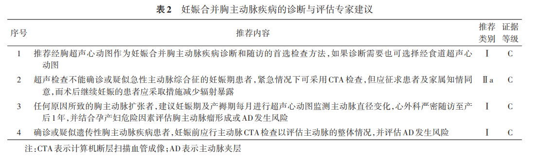
Experts' recommendations on diagnosis and assessment are shown in Table 2.
VI. Treatment and Management of Pregnancy Complicated by Thoracic Aortic Disease
01 Thoracic Aortic Dilation or Aneurysm
1. Complicated by Hereditary Connective Tissue Disorders
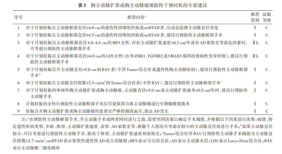
These are the leading cause of pregnancy-related thoracic aortic disease, with MFS being the most common (50%–68%). For MFS women planning pregnancy, if the aortic root diameter is 4.0–4.4 cm, especially with a growth rate ≥ 0.3 cm/year or family history, prophylactic aortic surgery may be considered preconceptionally. If the diameter is ≥ 4.5 cm, prophylactic surgery is routinely recommended before pregnancy.
For other syndromes (LDS, vEDS, Turner), prophylactic surgery should be considered when the ascending aortic diameter ≥ 4.5 cm, or the diameter index (diameter/BSA) ≥ 2.5 cm/m². The long-term rate of adverse aortic events after prior aortic root surgery is 21.4%.
2. Complicated by Bicuspid Aortic Valve (BAV)
About 50% of BAV patients have concomitant ascending aortic dilation. While the AD risk is lower than in MFS, it increases significantly when the ascending aorta reaches ≥ 5.0 cm (mWHO Risk IV), warranting consideration for prophylactic surgery.
3. Simple Thoracic Aortic Dilation or Aneurysm
After excluding hereditary causes, prophylactic surgery should be considered if the ascending aorta is ≥ 5.0 cm. Rapid expansion (≥ 0.5 cm/year) is also an indication. Decision-making requires comprehensive MDT evaluation considering genetics, mutation type, expansion rate, and family history.
Experts' recommendations on prophylactic timing are shown in Table 3.
4. Mode of Delivery
Evidence comparing delivery modes is lacking. Recommendations are:
Diameter < 4.0 cm: Vaginal delivery under strict monitoring.
Diameter ≥ 4.5 cm or prior AD: Cesarean section (C-section).
Diameter 4.0–4.4 cm: MDT evaluation required; C-section or assisted vaginal delivery with epidural analgesia is preferred.
Regardless of the mode, risks must be fully explained, and delivery should ideally occur under cardiac surgical supervision.
Experts' recommendations on delivery mode are shown in Table 4.

02 Aortic Dissection (AD)
1. Basic Principles
Basic management aligns with non-pregnant protocols: analgesia, heart rate control, and blood pressure control to reduce shear stress. Opioids are used for pain relief. Targets: Systolic BP 100–120 mmHg, Heart Rate 60–80 bpm. ACE inhibitors and ARBs are contraindicated. Refer to hypertension guidelines for specific regimens.
2. Surgical Intervention Strategy
Emergency green channels are mandatory. Surgical strategy depends on gestational age, dissection type, and fetal status, requiring informed consent.
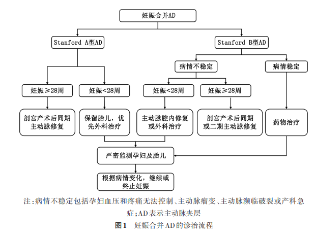
(1) Stanford Type A AD: Extremely life-threatening. The primary goal is saving the mother, followed by fetal salvage.
Gestational Age < 28 weeks:Maternal survival is prioritized. Fetal loss is likely, especially < 26 weeks. If the patient insists on continuing pregnancy post-surgery, strict fetal monitoring is required. For 26–28 weeks (periviable), C-section followed by aortic surgery may be considered to save the neonate.
Late Gestation:C-section followed by aortic surgery is recommended.
Intraoperative Measures:Femoral + axillary artery cannulation for perfusion; avoid deep hypothermic circulatory arrest if possible; prioritize valve-sparing or bioprosthetic root replacement; minimize CPB time; use antegrade cerebral perfusion.
Arch Reconstruction:Balancing simplicity (lower risk to fetus) vs. radicality (Sun's procedure, lower reoperation rate but longer CPB/arrest time). Decision based on maternal safety first.
Post-C-section Surgery:No fetal constraints; standard non-pregnant protocols apply. Manage postpartum hemorrhage risk (uterine balloon tamponade, low-dose oxytocin; avoid methylergometrine, carboprost).
Experts' recommendations are shown in Tables 5 & 6.
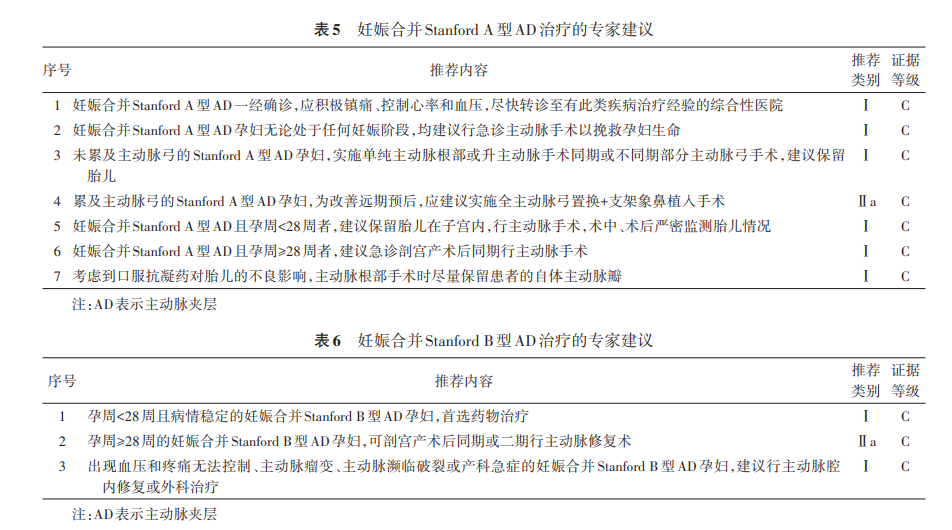
(2) Stanford Type B AD: Less immediately lethal than Type A. Medical therapy (rate/blood pressure control) is first-line.
< 28 weeks:Emergency intervention (TEVAR or surgery) if refractory pain/hypertension, impending rupture, or obstetric emergencies.
≥ 28 weeks:Consider fetal preservation or C-section, followed by staged or simultaneous aortic intervention.
TEVAR in late pregnancy/postpartum shows good mid-term results but requires minimizing radiation.
03 Cardiopulmonary Bypass (CPB) and Thoracic Aortic Surgery During Pregnancy
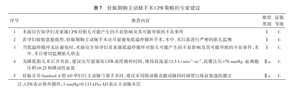
CPB poses significant risks to the fetus (hypoxia, miscarriage, etc.) due to reduced uteroplacental perfusion caused by low perfusion pressure, hemodilution, non-pulsatile flow, and hypothermia. Recommendations: Minimize CPB time; maintain high flow (>2.5 L·min⁻¹·m⁻²), high MAP (>70 mmHg), Hct >0.25, and pulsatile flow if possible. Mild hypothermia (28.1–34.0°C) may be tolerated, but deeper hypothermia for arch surgery severely compromises fetal survival. Decisions regarding circulatory arrest require balancing maternal/fetal factors and institutional capability.
Experts' recommendations are shown in Table 7.
04 Perioperative Anesthetic Management
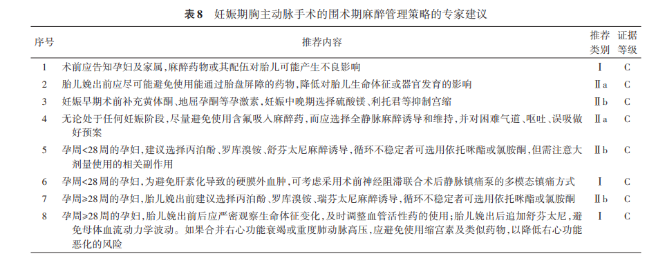
Anesthesia must consider effects on both mother and fetus. Risks are highest in the first trimester. Multimodal analgesia is recommended to reduce stress and prevent preterm labor. Drug selection must be precise regarding dosage and depth of anesthesia. Managing the difficult airway and transitioning between C-section and aortic surgery anesthesia are key challenges.
Experts' recommendations are shown in Table 8.
VII. Pregnancy Guidance
01 Genetic Counseling
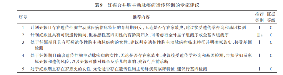
Thoracic aortic disease is closely linked to MFS, nsHTAD, LDS, and vEDS. Eleven genes are highly associated: FBN1, LOX, COL3A1, TGFBR1, TGFBR2, SMAD3, TGFB2, ACTA2, MYH11, MYLK,and PRKG1. 6–8% of hereditary cases harbor mutations without classic phenotypes. PGT-M (Preimplantation Genetic Testing for Monogenic disorders) is recommended for couples carrying known mutations. Amniocentesis/chorionic villus sampling is available for fetal screening. Women at mWHO ≥ II–III risk should undergo Ghent criteria evaluation, genetic counseling, and testing per the Expert Consensus on Genetic Testing and Clinical Diagnosis of Hereditary Thoracic Aortic Aneurysm/Dissection.
Experts' recommendations are shown in Table 9.
02 Guidance for Subsequent Pregnancies
Despite prophylactic surgery, residual risk remains, especially in LDS. Multidisciplinary preconception assessment is mandatory. PGT-M is an option. Reliable contraception is advised for those not desiring pregnancy; estrogen/progestin contraceptives should be avoided. Long-acting reversible contraception is preferred.
Absolute Contraindications to Pregnancy (Termination Recommended):
MFS: Aortic diameter ≥ 4.5 cm
BAV: Aortic diameter ≥ 5.0 cm
Turner Syndrome: Aortic size index ≥ 2.5 cm/m²
vEDS
Uncorrected severe coarctation of the aorta
Thoracic aneurysm ≥ 5.0 cm
History of AD or residual dissection with progressive dilation
VIII. Monitoring and Follow-up
All patients require close follow-up regardless of surgical intervention or pregnancy outcome.
Non-surgical patients: Monthly echocardiography during pregnancy and puerperium; monitor until 1 year postpartum. Increase frequency if high risk.
Surgical patients continuing pregnancy: Monthly fetal ultrasound until delivery.
Post-surgical/terminated patients: Standard non-pregnant follow-up protocols.
Aggressive control of risk factors is essential: BP < 120/80 mmHg, HR 60–80 bpm. Beta-blockers are recommended, especially for MFS, to slow dilation, but fetal growth must be monitored.
Breastfeeding decisions require MDT evaluation. It is contraindicated in patients with cardiac insufficiency or heart failure. Refer to Expert Consensus on the Diagnosis and Treatment of Pregnancy Complicated by Heart Disease (2016).
Source: Thoracic Aorta Committee of the Chinese Medical Doctor Association Society of Cardiovascular Surgery, Maternal-Fetal Critical Care Committee of the China Health Promotion Association for Science and Technology of Human Health, Cardiac Surgery Branch of the Beijing Medical Association. Expert Consensus on the Diagnosis and Treatment of Pregnancy Complicated by Thoracic Aortic Disease (2026). Chinese Journal of Obstetrics and Gynecology, 2026, 61(03): 177-189. DOI: 10.3760/cma.j.cn112141-20251012-00478
Editor: Lily