





















The ovarian reserve directly determines a woman's reproductive potential, age at menopause, and the risk of reproductive-related diseases. Textbooks and mainstream thought have long held that the peak number of germ cells in the female fetus reaches 7 million during the second trimester, sharply declining to 1–2 million at birth, representing a gestational loss of over 70%. This conclusion stems from the classic 1963 study by Baker and has been used ever since.
However, a systematic review published in Human Reproduction Update(2025) re-evaluated all quantitative studies on human fetal/newborn ovaries from 1953 to 2025. It revisited the classic understanding of the ovarian reserve during the human fetal-neonatal period, pointing out that the long-standing narrative of a "mid-gestation germ cell peak followed by massive pre-birth depletion" lacks robust evidentiary support. This provides a brand-new, evidence-based perspective for basic research on ovarian development and clinical reproductive assessment.
Source: Human Reproduction Update
01 Core Concepts and the Mainstream Narrative
1. Definition of Ovarian Reserve
It refers to the total volume of Non-Growing Primordial Follicles (NGF) within the bilateral ovaries. The level of this reserve at birth is directly linked to key reproductive outcomes such as age at menopause, premature ovarian insufficiency (POI), and the prognosis of assisted reproduction.
2. The Classic Mainstream Narrative
It is believed that the peak number of germ cells in the bilateral ovaries of a human female reaches 7 million during the second trimester. After massive apoptosis, only 1–2 million remain at birth, with the rate of germ cell depletion in the latter half of pregnancy reaching 70–85%. This conclusion has been widely incorporated into textbooks, guidelines, and public health content.
3. Origin of the Narrative
The core basis is the 1963 study by Baker, which has long served as the "gold standard" reference for ovarian reserve quantification.

Source: Human Reproduction Update
02 Key Research Basis: Re-analysis of Evidence
This review searched the literature up to September 2025, integrating seven studies from 1953 to 2011 involving 139 human embryo-fetal-newborn ovarian samples. It compared different counting methods and data outcomes:
1. Core Data from Baker (1963)
At 20 weeks of gestation: 6.8 million germ cells in bilateral ovaries (20% atretic).
At birth: 2 million germ cells in bilateral ovaries (50% atretic).
Study Defects: Extremely small sample size (only 2 ovaries per time point); delayed tissue fixation leading to cytolysis; volume estimation methods prone to overestimation; inaccurate identification of apoptotic cells; and a lack of independent verification for over 60 years.
2. Wallace & Kelsey (2010) Mathematical Model
Modeling integrated 325 ovarian samples from 8 studies.
Peak unilateral ovarian NGF at mid-gestation: 300,000; at birth: 295,000.
After bilateral conversion: 600,000 at mid-gestation and 590,000 at birth, indicating no massive germ cell loss.
3. Modern Stereological Study Data
At 19–20 weeks of gestation: Mean bilateral ovarian germ cells = 323,000.
At birth: Mean bilateral ovarian germ cells = 669,000.
Results varied widely across different studies (ranging from thousands to 20 million), with no data supporting Baker’s million-level quantities.
03 Core Findings: The Truth About Germ Cell Quantity and Apoptosis
1. Germ Cell Quantity: Classic Data Severely Overestimated
Estimates of fetal-newborn ovarian germ cells are far lower than the traditional 7 million to 2 million range; modern reliable data are approximately 10 times lower than Baker’s results.
Only 1% of newborns have ≥1 million NGFs in their bilateral ovaries, contradicting the belief that the average at birth is 1–2 million.
2. Germ Cell Apoptosis: No Evidence of Massive Depletion
Classic Belief: 20% apoptosis at mid-gestation, 50% at birth, totaling 70–85% depletion.
Modern Evidence: Detection of apoptosis markers (TUNEL, caspase-3, Bax) shows that the fetal germ cell apoptosis rate is mostly <5%, with the highest reported rate being only about 20% (subjectively determined in a single study).
High-quality histological sections show no widespread germ cell apoptosis, failing to support the conclusion that "millions of germ cells are lost before birth."
3. Correlation Between Ovarian Volume and Reserve
Adult Women: Strong correlation between ovarian volume and NGF count.
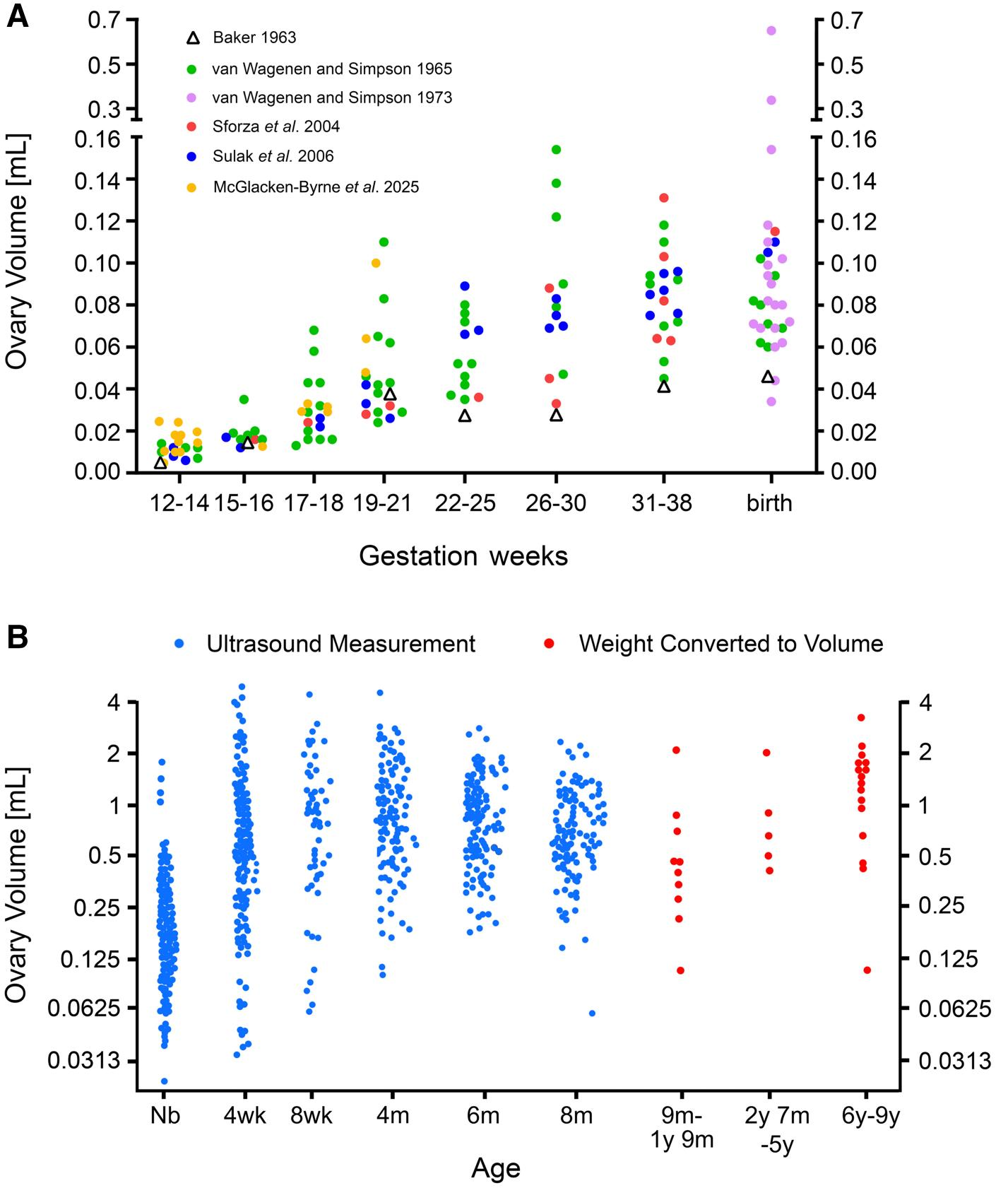
Fetus-Newborn: Individual differences in ovarian volume can reach 50-fold; the morphology is irregular and shows no clear correlation with germ cell quantity.
Although ovarian volume increases significantly from mid-gestation to birth, the number of germ cells does not drop sharply.
Source: Human Reproduction Update
04 Reasons for Discrepancies and Methodological Issues
1. Differences in Counting Methods
(1) Volume Estimation/Model Correction (used by Baker): Prone to overestimation bias due to susceptibility to ovarian shape and cell distribution.
(2) Unbiased Design Stereology/Optical Dissector (Modern Mainstream): Not affected by ovarian shape or volume, yielding more accurate results.
2. Cell Identification Errors
Meiotic oocytes and apoptotic cells have similar morphology, making it difficult for traditional sections to distinguish them precisely, leading to misjudgment of the number of atretic cells.
3. Sample and Technical Limitations
Early studies had extremely small sample sizes and non-standardized tissue processing. Modern studies still suffer from insufficient sample sizes and high data heterogeneity.
05 Clinical and Research Implications
1. Reproductive Clinical Assessment
The level of ovarian reserve at birth directly affects clinical decisions regarding POI, early menopause, chemotherapy-induced ovarian damage, and IVF prognosis.
The overestimated traditional认知 (cognition) of "1–2 million at birth" needs correction to optimize fertility counseling and risk assessment.
2. Basic Research Directions
Large-sample, standardized, unbiased stereological studies are needed to systematically quantify total germ cell numbers and the ratio of surviving/apoptotic cells at various stages of pregnancy.
3. Revision of Education and Public Awareness
The narrative of "7 million → 1–2 million" circulating in textbooks and online should be updated to conclusions supported by scientific evidence.
06 Research Limitations and Guidance
1. Limitations
Overall small sample sizes in included studies and high heterogeneity in some data.
Insufficient paired data synchronizing fetal-newborn ovarian volume with germ cell quantity.
The molecular mechanisms of germ cell apoptosis and precise quantification still require in-depth exploration.
2. Guidance for Research and Clinical Practice
Abandon the rigid reliance on Baker’s study and base assessments on modern unbiased quantitative data.
Ovarian reserve assessment must prioritize individual variation rather than applying a uniform mean.
The genetic regulatory mechanisms of germ cell development and apoptosis are key future research directions.
The classic narrative that millions of germ cells form in the human fetal ovary during the second trimester and undergo 70–85% massive apoptosis before birth lacks reliable scientific support. Modern research confirms that the number of fetal-newborn ovarian germ cells is far lower than traditionally believed, the apoptosis rate is extremely low, and there is no massive depletion. The process of establishing the ovarian reserve, its quantitative levels, and individual differences still require clarification through large-scale, standardized studies. This conclusion will reshape the understanding of human ovarian development and optimize the direction of clinical assessment and basic research in reproductive health.
Source: Jeffrey B Kerr, Raymond J Rodgers, The human ovarian reserve: the narrative and the science, Human Reproduction Update, 2025;, dmaf031, https://doi.org/10.1093/humupd/dmaf031
Editor: Lily